

Full Length Research Article
Detection of Novel L-arginase Gene Sequences from Pseudomonas aeruginosa in Soil and Sewage Samples
Zainab Abbas Abd1*, Aqeel Mohammed Majeed Al- Ezee2, Zaid Raad Abbas2
Adv. life sci., vol. 12, no. 2, pp. 291-295, May 2025
*- Corresponding Author: Zainab Abbas Abd (Email: zainab19@uomustansiriyah.edu.iq)
Authors' Affiliations
2. Department of Microbiology, College of Science, Mustansiriyah University – Iraq
[Date Received: 20/10/2024; Date Revised: 05/01/2025; Date Published: 22/03/2025]
Abstract![]()
Introduction
Methods
Results
Discussion
References
Abstract
Background: Pseudomonas aeruginosa is known for its flexibility and importance as both a pathogen and a model organism for genetic studies. This investigation was conducted to detect L-arginase enzyme gene sequences from P. aeruginosa in soil and sewage samples.
Methods: Fifty-two (52) soil and sewage samples were collected from different regions in Iraq for six months, from November 2022 to April 4, 2023. Pseudomonas aeruginosa was isolated by culturing samples on nutrient medium and McConkey agar medium. Gram stains and biochemical tests were performed to identify the isolates, and the VITEK 2 system was used to confirm the identity of P. aeruginosa. DNA was extracted from the P. aeruginosa isolates and used for molecular identification by amplifying and sequencing the 16S rRNA gene. Also, the L-arginase gene sequences were amplified using the PARG1 and PARG2 primers and sequenced.
Results: The results showed that P. aeruginosa isolates from sewage water (18 [34.6%]) were higher than those obtained from the soil (9 [53.57%]). The PCR technique confirmed the identity of P. aeruginosa. Furthermore, the PCR technique revealed that the novel L-arginase enzyme gene sequence was 300 bp in size.
Conclusion: The present study's findings revealed the size and genetic sequence of the novel L-arginase enzyme from P. aeruginosa isolates derived from soil and wastewater samples. This research is considered a crucial step toward understanding the genetic structure and functions of arginase in P. aeruginosa, providing insights for future scientific investigations.
Keywords: Gene sequences; L-arginase; PCR; Pseudomonas aeruginosa
Introduction![]()
Pseudomonas aeruginosa is a versatile Gram-negative bacterium known for its adaptability and significance as both a pathogen and a model organism for genetic studies. Its genetic manipulation has provided valuable insights into bacterial gene expression and regulation. One important aspect of P. aeruginosa gene construction is identifying and isolating the target gene of interest. This gene can be obtained from P. aeruginosa, other organisms, or synthesized de novo. Polymerase chain reaction (PCR) and genome sequencing techniques facilitate gene acquisition. Pseudomonas aeruginosa gene construction begins with identifying and isolating the target gene of interest. Once the gene is isolated, various molecular biology techniques can be employed to manipulate it [1]. Selecting the right genetic components is essential for gene expression in P. aeruginosa gene construction. This includes choosing appropriate promoters, ribosome binding sites, and transcriptional terminators to control gene expression levels and ensure proper regulation.
Researchers often optimize these elements to achieve the desired gene expression patterns. Once the gene is successfully integrated or introduced into P. aeruginosa, further characterization and analysis are performed to verify its functionality and assess the phenotypic effects. This involves applying techniques, such as PCR, gene expression profiling, protein analysis, or functional assays to verify gene functionality and assess phenotypic effects [2, 3].
L-arginase is an enzyme that catalyzes the hydrolysis of L-arginine into L-ornithine and urea. While L-arginase is commonly found in many organisms, including bacteria, its presence and role in P.aeruginosa has been the subject of investigation. This bacterium has been reported to possess L-arginase activity.
It is known that L-arginase in P. aeruginosa is involved in numerous physiological activities, including nitrogen metabolism and adaptation to different environmental conditions. Also, it has been suggested that L-arginase activity in P. aeruginosa contributes to its virulence and pathogenesis [4].
L-arginase activity in P. aeruginosa has been associated with regulating the production of secondary metabolites and quorum-sensing molecules. The expression of several virulence factors in P. aeruginosa has been proposed to be influenced by L-arginase [5]. Pseudomonas aeruginosa has been detected in sewage treatment plants. Studies have investigated its prevalence, antibiotic resistance, and virulence factors in this context. Poursina et al. [6], carried out an investigation on P.aeruginosa in sewage treatment plants and reported on the prevalence, antibiotic resistance patterns, and virulence factors of P.aeruginosa strains isolated from these environments. This investigation aimed to detect novel L-arginase enzyme gene sequences from P.aeruginosa in soil and sewage samples obtained from various locations in Iraq.
Methods![]()
Collection of samples
Fifty-two soil and sewage samples were collected for six months, from November 2022 to April 4, 2023, from various locations in Iraq. The samples were carefully transported to the laboratory for further analysis.
Laboratory examination of collected samples
The samples were cultured on a medium, such as nutrient medium and McConkey agar medium to isolate Pseudomonas aeruginosa. After that, the samples were stained with Gram stain, then biochemical tests were performed to identify the isolates. The identity of all the P. aeruginosa isolates was confirmed using VITEK 2 (BioMérieux).
Identification of Pseudomonas aeruginosa using the polymerase chain reaction
DNA was extracted from the P. aeruginosa isolates and subjected to PCR-amplification to detect the PA-SS (16SrRNA) gene to identify P.aeruginosa species.
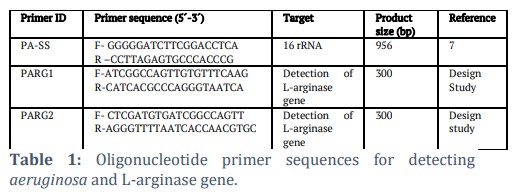
The PCR mix was made in a total volume of 25 μl comprising oligonucleotide primers (forward and reverse) and Quick-Load® Taq 2X Master Mix, which was liquefied at room temperature. Table 1 shows the nucleotide sequences of the oligonucleotide primers used for amplification.
Detection of L-arginase gene
The design of primers (Table 1)to detect the gene encoding the L-arginase enzyme was carried out in a scientific laboratory at Al-Mustansiriyah University, Iraq.
The amplification conditions for the primers were optimized. The PCR to amplify the L-arginase gene was performed in a thermal cycler with the following conditions: 95oC for 2 min (initial denaturation), followed by 35 cycles of 94oC for 20 sec (denaturation), 58oC for 20 sec (annealing), 72oC for 40 sec (extension), and one cycle of 72oC for 1 min (final extension).
Results![]()
Fifty-two bacterial isolates were obtained from different agricultural soils and sewage water, with a distribution of 28 (53.8%) from soil samples and 24 (46.2%) from sewage samples, as presented in Table2.
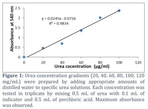
The regular curve of urea in the present study showed that urea production by arginase was very high with a value of 110 µg/ml (Figure 1).
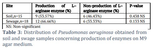
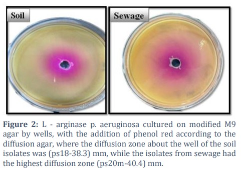
Out of 33 (63.46%) isolates of P. aeruginosa from soil and sewagesamples,only9 (53.57%) of the soil isolates produced the L–arginase in M9 media, while 12 (66.66%)isolates from the sewage water produced the L–arginase in M9 media (Table3 and Figure 2).
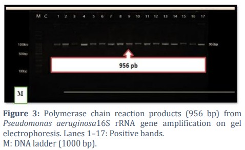
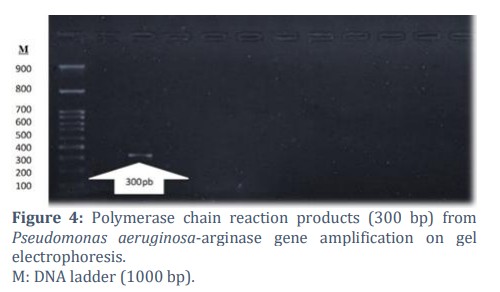
The results obtained for the molecular identification of P. aeruginosa, and detection of L-arginase gene sequences are shown in Figures 3 and 4, respectively. PCR-product fragment sizes of 956 and 300 bp were obtained after the amplification of the rRNA and L-arginase genes, respectively.
Figures & Tables
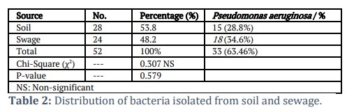
The present study's findings revealed no substantial differences in the number of isolates from soil and sewage. However, the number of isolates in the soil was more than in sewage water. This is because bacteria in the soil are more than other microorganisms due to their ability to replicate easily. The observations made in the present study are consistent with previous findings[8,9]. In addition, sewage water provides an ideal environment for the growth of pathogenic and non-pathogenic bacteria, because it contains many elements necessary for the growth and reproduction of these bacterial species, as reported by Mohammed [10].
In the present study, arginase produced a relatively high amount of urea (110 µg/ml). The high production of urea observed during the assay of the L-arginase enzyme by M9 enhanced the color change from yellow to pink (Figure 2). This is crucial in the conversion of ammonia to urea in the urea cycle. Furthermore, the activity of the enzyme can be stimulated by measuring the volume of the enzyme (ml) and the amount of urea released during the enzyme's action. This finding highlights the significance of arginase in maintaining ammonia homeostasis and suggests that enzymatic activity is functioning efficiently in the given context, which is in agreement with previous reports that have demonstrated the significance of arginase in the regulation of levels of urea production [11]. The measurement of urea concentration provides a valuable indicator of arginase activity and the proper functioning of the urea cycle. Also, Lee and coworkers found that an increased concentration of urea was associated with increased metabolism of arginine [12].
The use of the PCR method in the identification of P.aeruginosa is a commonly employed molecular technique in research and clinical settings. The procedure allows for the amplification of specific regions of the bacterial DNA, enabling sensitive and specific detection of the test organism. This procedure was used by Li et al. (2011) whose study focused on the rapid detection of P. aeruginosa in medical samples utilizing real-time PCR. The researchers highlighted the benefits of the RT-PCR, including its sensitivity, specificity, and quick turnaround time for detecting P. aeruginosa infections in a clinical laboratory setting. As far as we are aware, the present study presents the first report on the extraction of the arginase gene from P.aeruginosa. This finding contributes to the understanding of the genetic repertoire of P.aeruginosa and expands our knowledge of the functional genes present in this bacterium.
The arginase gene is made up of a 300bp DNA fragment and encodes the enzyme arginase. The enzyme catalyzes the hydrolysis of arginine to ornithine and urea, contributing to various physiological processes and metabolic pathways in the cells [11]. Pseudomonas aeruginosa is a versatile and adaptable bacterium known for its metabolic diversity and ability to thrive in various environments [14]. The identification and characterization of the arginase gene in P.aeruginosa provides insights into its potential role in arginine metabolism and the adaptability of this bacterium to different ecological niches.
The discovery of the arginase gene in P.aeruginosa has significant implications. It opens avenues for further research into the functional properties of the enzyme and its potential applications. Understanding the regulation and expression of the arginase gene can provide insights into the metabolic pathways involved in arginine utilization by P.aeruginosa. Furthermore, the arginase gene may have implications for pathogenicity and virulence. Pseudomonas aeruginosa is an opportunistic pathogen associated with numerous infections, particularly in immune-compromised individuals [15]. The arginase enzyme can influence the availability of arginine, an essential amino acid for immune cells, and potentially impact host-pathogen interactions and disease progression.
The study’s findings revealed a novel pathway, presenting an opportunity for scientific exploration and the potential for leveraging this enzyme's capabilities in bacterial growth research. Also, the present study highlights the possibility of harnessing arginase from natural sources, offering a new approach to inquiring about its applications in scientific investigations. This research signifies a breakthrough in genetic manipulation and opens up promising prospects for future studies in microbiology and related disciplines.
Acknowledgement
The authors much appreciate the management of Al-Mustansiriyah University for their help in carrying out this investigation.
Author Contributions
The conceptualization of this study was done by ZA, AM carried out soil and sewage sample collection. ZA conducted the laboratory analysis. ZA prepared the draft manuscript, which AM and ZA proofread. Both authors agreed to send the manuscript to Advancements in Life Science journal for publication consideration.
The authors declare that there is no conflict of interest.![]()
References
- Tatusova T, DiCuccio M, Badretdin A, Chetvernin V, Nawrocki EP, et al. NCBI prokaryotic genome annotation pipeline. Nucleic Acid Research, (2016); 44(14): 6614-6624.
- Limoli DH, Whitfield GB, Kitao T, Ivey ML, Davis MR Jr, et al. Pseudomonas aeruginosa alginate overproduction promotes coexistence with Staphylococcus aureus in a model of cystic fibrosis respiratory infection. Molecular Biology and Microbiology, (2017); 8(2): e00186-17.
- Liberati NT, Urbach JM, Miyata S, Lee DG, Drenkard E, et al. An ordered, nonredundant library of Pseudomonas aeruginosa strain PA14 transposon insertion mutants. The Proceedings of the National Academy of Sciences,(2006); 103(8): 2833-2838.
- Tortoli E. Commentary on phylogenomics and comparative genomic studies robustly support division of the genus Mycobacterium into an emended genus Mycobacterium and four novel genera. Frontier of Microbiology, (2018); 9: 2065.
- Scribani RC, Barrientos-Moreno L, Paone A, Cutruzzolà F, Paiardini A, et al. Nutrient sensing and biofilm modulation: the example of L-arginine in Pseudomonas. International Journal of Molecular Science, (2022); 23(8): 4386.
- Poursina S, Ahmadi M, Fazeli F, Ariaii P. Assessment of virulence factors and antimicrobial resistance among the Pseudomonas aeruginosa strains isolated from animal meat and carcass samples. Journal of Veterinary Medical Science, (2023); 9(1): 315-325.
- Spilker T, Coenye T, Vandamme P, LiPuma JJ. PCR-based assay for differentiation of Pseudomonas aeruginosa from other Pseudomonas species recovered from cystic fibrosis patients. Journal of Clinical Microbiology, (2004); 42(5): 2074-2079.
- Ingham ER. Soil Biology Primer, Chapter 4: Soil Fungi. In: Soil and Water Conservation Society (Ed: Ankeny IA), (2009); pp22-23.
- Alown F, Alsharidah A, Shamsah S. Genotypic characterization of soil bacteria in the Umm Al-Namil Island, Kuwait. Saudi Journal of Biological Sciences. 2021; 28(7): 3847-3854.
- Todd ECD. Waterborne Diseases and Wastewater Treatment in Iraq. Journal of Food Protection. 2024; 87(1): 100204.
- Morris SM. Arginases and arginine deficiency syndromes. Current Opinion in Clinical Nutrition and Metabolic Care, (2018); 21(1): 15-21.
- Li Z, Wang L, Ren Y. Huang Y, Liu W,et al. Arginase: shedding light on the mechanisms and opportunities in cardiovascular diseases. Cell Death Discovery, (2022); 8(1): 413.
- Lee CS, Wetzel K, Buckley T, Wozniak D, Lee J. Rapid and sensitive detection of Pseudomonas aeruginosa in chlorinated water and aerosols targeting gyrB gene using real-time PCR. Journal of Applied Microbiology, (2011); 111(4): 893-903.
- Harrison E, Lycett S, Koskella B, Perron GG. The emergence of plasmid stability under non-selective conditions maintains antibiotic resistance in the absence of resistance-driving plasmids. Molecular Biology and Evolution, (2021); 38(1): 45-55.
- Aslantaş Ö, Türkyilmaz S, Keskin O, Güllü Yücetepe A, Büyükaltay K. Molecular Characterization of Pseudomonas aeruginosa Isolated From Clinical Bovine Mastitis Cases Klinik İnek Mastitis Vakalarından İzole Edilen Pseudomonas aeruginosa Suşlarının Moleküler Karakterizasyonu. Kafkas Universitesi Veteriner Fakultesi Dergisi. (2022) ;28(6): 747-759.
This work is licensed under a Creative Commons Attribution-Non Commercial 4.0 International License. To read the copy of this license please visit: https://creativecommons.org/licenses/by-nc/4.0


