

Full Length Research Article
Clinical study of common bacterial, fungal and parasitic skin diseases in cats
Hassanin H. N. Alautaish1*, Hussein Ali Naji1, Zainab Abudal Hussein Saud2, Baneen Ghanim Ghalib1
Adv. life sci., vol. 11, no. 3, pp. 580-584, August 2024
*- Corresponding Author: Hassanin H. N. Alautaish (hussein.naji@uobasrah.edu.iq)
Authors' Affiliations
2. Department of Public Health, College of Veterinary Medicine, University of Basrah. Basra – Iraq
[Date Received: 10/05/2023; Date Revised: 27/02/2024; Date Published: 10/07/2024]
Editorial Expression of Concern:
18 May 2025: Following publication of this paper, the internal audit (consequent to concerns on quality raised by Web of Science) notified Advancements in Life Sciences about problems in use of English language. By this Editorial Expression of Concern, we alert the scientific community as we address the errors.
Editorial Note:
31 May 2025: You are viewing the latest version of this article having minor corrections related to the use of English language. Expression of concern is hereby revoked.
Abstract![]()
Introduction
Methods
Results
Discussion
References
Abstract
Background: The skin is a large, metabolically active organ that requires significant nutrients and protein in its body. The study pointed high significant infection with fungal, scabies, alopecia, bacterial and wounds in the white color cats more than the others because it is represented the better color choice for the families then the orange and grey colors while the black color is the less in number of domestic cats. The aim of present study was investigated of some skin disease in cats in Basrah, Iraq.
Methods: The present study includes two hundred animals of both sexes with different ages, colors, weights and sizes were studied clinically in Basrah veterinary hospital and the private veterinary clinics in Basrah. The study extended from October 2022 to March 2023 during the winter season. Clinical signs were reported carefully, and the initial diagnosis of skin diseases was confirmed by apparent lesions on the skin, the skin samples of the cases were collected for laboratory diagnosis in veterinary medicine college ̸ Basrah University and Basrah veterinary hospital laboratories to confirmed diagnosis.
Results: Clinical signs were reported carefully and the initial diagnosis of skin diseases. The two hundred sample cases were divided according to cases in Basrah veterinary hospital and Basrah private clinic: 60 Cases fungal infection, 40 cases scabies, 10 case alopecia, 50 cases of bacterial infection and 40 cases of wound.
Conclusion: The present study shows the fungal diseases were common causes for skin diseases in Cats follow by Bacterial, Parasitic and Alopecia respectively.
Keywords: Cat infection; Skin diseases in cat; Scabies; Fungal disease
Introduction![]()
The skin is a large, metabolically active organ that requires significant nutrients and protein in its body. The condition of its skin and coat may be impacted by variations in its nutritional supply [1,2]. In three ways, dietary factors may be significant in the development and management of skin disease: nutritional imbalance or deficiency, therapeutic use of nutritional supplements, and dietary sensitivity [3].
Scabies in cats is a highly contagious disease that caused by a tiny mite named Notoedres cati and Sarcoptes scabiei [4]. Scabies burrow in the skin and cause a crusty dermatose, primarily in sparsely hairy areas, including the head, neck, axillae, groin and tail [5]. Ordinary scabies present at burrowing site as popular or vesicular lesions, and generalized allergic rash accompanied by intense itching [6]. Therefore, scabies in cats must be treated to prevent thickening and skin folding which formed due to crust [7].
Dermatophytosis, it is the most frequent fungal infection in cats and one of the most significant infectious skin illnesses, usually caused by Microsporum canis. Many adult cats have the disease asymptomatically. Severe clinical symptoms are primarily observed in immunocompromised adults or kittens. In shelters or batteries, the disease may be endemic and poor cleanliness is a predisposing factor. The environmental lifespan of dermatophyte-produced infectious arthrospores is approximately one year. They can be spread by coming into touch with healthy or sick cats, as well as by dust, clothing, brushes, and other foreign objects. It is common to have erythematous margins, desquamation, and circular alopecia surrounding center healing (also known as "ringworm"). This is a self-limiting illness that merely causes scaling and hair loss in many cats. When cats are immunosuppressed, the result could [8].
The bacterial infections could be a source of disease or illness if the pathogenic bacteria are present in animals [9]. Bacterial skin infection in the cat is present and sub- cutaneous abscesses are the most common forms of infection usually due to bite wounds. Feline superficial and deep infections are almost always associated with other underlying disease processes such as metabolic or immunological abnormalities. The main pathogen in superficial infections is Staphylococcus intermedius. In deep pyoderma many different aerobic and anaerobic bacteria including Pasteurella multocida, beta-hemolytic streptococci, Actinomyces spp. Bacteroides spp. and Fusobacterium spp. can be identified [10].
Skin wound developed to ulcers in cats are defects in the surface layers of a feline’s skin. The majority of skin wound are the result of trauma from an outside source but can also be linked to certain varieties of disease [11].
Hair loss that affects the ventral, lateral, perineal, and dorsal parts of a cat's trunk and typically occurs in a symmetric manner is known as focal or generalized alopecia. This could be caused by an inability to produce enough hair coat, excessive hair loss as a result of self-trauma, or excessive complete hair shedding. The most frequent cause of hair loss is self-trauma, which is especially linked to dermatitis caused by flea allergies [12].
The aim of present study was investigated of some skin disease in cats in Basrah, Iraq.
Methods![]()
The study conducted on the main four domestic breeds of cats in Basrah_ Iraq. The present study include two hundred animals of both sexes with different ages, colors, weights and sizes were studied clinically in Basrah veterinary hospital and the private veterinary clinics in Basrah. The study extended from October 2022 to March 2023 during the winter season. Clinical signs were reported carefully, and the initial diagnosis of skin diseases was confirmed by apparent lesions on the skin, such as desquamation, apparent ulcers, a change in skin color, or hair loss. Then; skin samples of the cases were collected for laboratory diagnosis in veterinary medicine college ̸ Basrah University and Basrah veterinary hospital laboratories to confirmed diagnosis.
All two hundred samples were checked under direct examination by microscope by 10X, And 40 X to identification of skin disease¢s causes such as fungi, bacteria, and parasite or any other causes such as nutritional causes and skin sensitivity.
Results![]()
The two hundred sample cases were divided according to cases in Basrah veterinary hospital and Basrah private clinic: 60 Cases fungal infection, 40 cases scabies, 10 case alopecia, 50 cases of bacterial infection and 40 cases of wound as illustrated in Table (1). There were no significant changes in body temperatures, heart and respiratory rats therefore it is ignored in the present study.
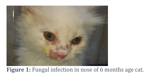
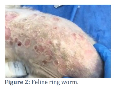
Fungal Infection: According to samples received from Basrah veterinary hospital and Basrah private clinic 60 cases which form 30% show the classic appearance of a round red ring that strikes a cat which give to fungal infection scientific name Feline Ringworm, where the lesions take close characteristic signs such as circle area of hair loss, broken and stubbly hair scaling skin, changes in hair or skin color, dermatitis in severe cases, excessive grooming, scratching, infected nail beds and sometime dandruff.
The common site of infection in the ears, mouth, nose respectively may be in other site such as nose, face, and front legs. The figures 1, and 2 show the lesion in some animals undergo of fungal infection.
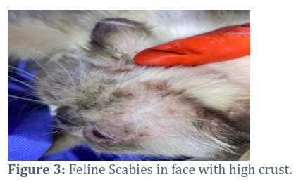
Scabies Infection: Forty cases which form 20% of the total reported cases were suffering from scabies infection with exhibited redness, rash, and severe itching associated with blood from the skin and hair loss in a large way and may be crusted on the edges of the infection area especially the ear and ear edges. The laboratory examination revealed to the Notoedric cat (Feline Scabies) infection, in addition to the occurrence of large ulceration in the skin and a rise in temperature. The common site of infection in all body part, nose and mouth this figure show infection received from hospital and private clinic figure (3) show these infections.
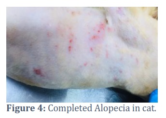
Alopecia: According to the reported cases; 10 affected animals from Basrah private clinic which appear a severe hair loses in some body parts developed partial or complete alopecia which occur due to systemic disease or skin sensitivity or uncleaned and dirty places in Figure (4), there cases reported 5% of studied cases.
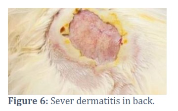
Bacterial Infection: Fifty cases which caused dermatitis characterized by scratching, red papules bump on the skin, scaly patches, itchiness, especially around the face and feet, frequent head shaking, thick, dark patches of skin, Patches of hair loss. The Common site of infection in head and back as in figure (5) where show severe dermatitis with bacterial infection confirmed by laboratories in Basrah veterinary hospital, these cases represented about 25% of total studied cases.
Wounds: Forty cases which caused by injures that appear a scratching, bleeding and frequent head shaking. The Common site of infection in all body part, these cases formed about 20% of total studied cases.
Figures & Tables

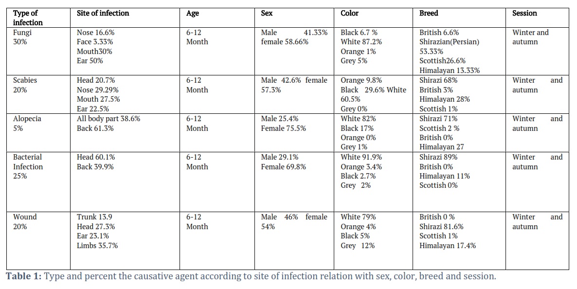
The study pointed high significant infection with fungal, scabies, alopecia, bacterial and wounds in the white color cats more than the others because it is represented the better color choice for the families then the orange and grey colors while the black color is the less in number of domestic cats. Moreover, the study reported the same results in the Persian breed which known locally as (Sherazin) more than the others due to the same reasons and the Himalayan breed occupied the second order then the Scottish and British breeds, all breeds can affect with the infections with different levels as explained in the results. Also, among all study subjects, females showed a higher percentage of infections because due to their greater numbers than males.
The age of infection ranged between 6 to 12 months, that age represent the vital age for cats to play and explorer the environment therefore it will be with direct contact to the contamination and the source of infections beside the incomplete immune system compared to the aged cats. This is corresponding with [13].
The season of infections play an important role in the infection, the study occurs in autumn and winter which are the period of study, where the moderate temperature the year in Basrah with low temperatures in some days of late December and January, with moist weather, which can be regarded as predisposing factor to induce these infections in addition the weather changes play very important role in the cleanliness of cat’s body because the owners avoid pathing their pets fearing of the illnesses that prepare for skin infection or changes. These results similar to that reported by [14].
Cat ringworm often manifests as a regular, circular alopecia accompanied by hair breaking, desquamation, and occasionally an erythematous perimeter and central healing. As stated by some patients exhibit erythema, pruritus, exudation, crusts, and peculiar, big alopecic [15]. he signs mainly reported in in ears and mouth it may be due the moist skin in this parts that is necessary fungal life.
One or more mite species, with varying incidence and toxicity, can cause scabies, a skin condition that is transmitted by direct contact with infected animals or contaminated settings. When an animal is attacked, it will become itchy, which will lower its body condition and result in financial losses. The nose has the highest infection rate because as it is the organ most frequently in contact with sources of infection these results agree with [16].
Lesions from dermatophytosis in cats are more pleomorphic. One or more partial alopecia patches with scaling and crusting, usually on the head or forelimbs, are considered classic lesions. Hyperpigmented lesions are possible [17]. Additionally, dermatophytosis can cause isolated, itchy lesions that resemble eosinophilic plaques or lesions that mimic miliary dermatitis. Given that diseased hairs are prone to breakage, long-haired cats may complain of having a poor hair coat or excessive shedding [18]. Although Persian and Himalayan cats are less likely to get kerions, they can develop subcutaneous nodular lesions known as dermatophytic pseudomycetoma, which are caused by dermatophytes. These lesions are frequently draining, consolidating nodules or exudative lesions. These lesions may be accompanied with visible tissue grains. Additionally, dermatophytic pseudomycetoma systemic lesions have been [19].
Alopecia is one of the most important lesion affected animals especially on the back and it is reported in all parts of the body as partial or complete lesion; these lesions may occur due to the bad nutrition or minerals deficiency such as Zink or Cupper deficiency and multivitamin deficiency such as Vitamin E and A deficiency, moreover it occur secondary to other skin lesions for example: allergy, pruritus, dermatosis and finally external parasitic invasion this explained by [20].
Erythema is a primary lesion of cats, pruritus and inflammation can result in self-induced alopecia, excoriation and secondary infections with papules, pustules and crusts [2]. Axillae, abdomen, distal extremities, inner pinnae and periocular, perioral and perianal regions are commonly affected [2]. Otitis externa is present in half of cats. Predilection sites differ from breed to breed but head is more affected as explained by [21].
The environment for regulated bacterial development is the skin. Numerous investigators have documented the intricate relationship between cutaneous infection and ambient and local variables, host immunity, organism adhesion, and virulence. Gram-positive bacteria that are present in the home include Corynebacterium sp., Micrococcus, and Staphylococcus. Skin pathogens Streptococcus pyogenes and Staphylococcus aureus are well-known. Bacteria must be able to stick to, develop on, and infiltrate the host in order for them to be considered pathogenic. Many virulence genes in bacteria enable them to flourish in these exclusive habitats. The infections of the skin that are brought on by S. aureus and S. pyogenes are impetigo and ecthyma. Cellulitis, necrotizing fasciitis, and erysipelas are examples of dermal infections. Furunculosis, carbunculosis, and folliculitis are all associated with the pilosebaceous unit. Additionally, the toxins that S. aureus and S. pyogenes produce [22].
Wound due to fighting and bad grooming or sequala of other dermatological infection with untreated cases such as a layer of crusts, dermatitis also it may be occurred due to excessive itching and pruritus in some cases.
Fungal infection is the primary skin disease in cats. Complete alopecia is less prevalent. The head is the most susceptible site for skin infections.
Conflict of Interest
The authors have no conflicts of interest to declare.
Conceptualization: HHNA, HAN, ZAS, BGG
Data curation: HHNA, HAN, ZAS
Investigation: HHNA, BGG
Methodology: HHNA, HAN, BGG
Project administration: HHNA, HAN
Supervision: HHNA, HAN
Writing Original draft: HHNA
Writing-review & editing: HHNA, HAN
Hassanin H. N. Alautaish (HHNA), Hussein Ali. Naji (HAN), Zainab AbudalHussein Saud (ZAS), Baneen Ghanim Ghalib (BGG)
![]() References
References
- Abdullhussain TH, Naji HA. Prevalence Study of Canine Infectious Hepatitis in Puppies and Adult Dogs in Basrsh Province, Iraq. World of Science: Journal on Modern Research Methodologies, (2023); 2(2): 30-39.
- Naji HA, Saleh WM, Saud ZA, Mhahal TR, Alhasson FA, et al. Prevalence of resistance and virulence genes in Escherichia coli isolates from diarrheic dogs. Iraqi Journal of Veterinary Sciences, (2023); 37(2): 355-361.
- Al-Autaish HH, Hasso SA. Identification of lyst gene in iraqi water buffaloes (Bubalus bubalis) with chediak-higashi syndrome by rflppcr method. Biochemical & Cellular Archives, (2020); 20(2): 5021-5027.
- Black SS, Abemethy TE, Tyler JW, Thomas MW, Garma‐Aviña A, et al. Intra‐abdominal dermatophytic pseudomycetoma in a Persian cat. Journal of veterinary internal medicine, (2001); 15(3): 245-248.
- Chiller K, Selkin BA, Murakawa GJ. Skin microflora and bacterial infections of the skin; 2001. Elsevier. pp. 170-174.
- Diesel A, DeBoer DJ. Serum allergen‐specific immunoglobulin E in atopic and healthy cats: comparison of a rapid screening immunoassay and complete‐panel analysis. Veterinary dermatology, (2011); 22(1): 39-45.
- Favrot C. Clinical signs and diagnosis of canine atopic dermatitis. In: 3. Congresso Latinoamericano de Dermatologia Veterinaria, Buenos Aires, Argentina, 26 – 27 November (2015); s.n.. https://doi.org/10.5167/uzh-116541
- Frymus T, Gruffydd-Jones T, Pennisi MG, Addie D, Belák S, et al. Dermatophytosis in cats: ABCD guidelines on prevention and management. Journal of feline medicine and surgery, (2013); 15(7): 598-604.
- Gedon NKY, Mueller RS. Atopic dermatitis in cats and dogs: a difficult disease for animals and owners. Clinical and translational allergy, (2018); 8(1): 1-12.
- Goldust M, Rezaee E, Raghifar R, Naghavi-Behzad M. Ivermectin vs. lindane in the treatment of scabies. Annals of parasitology, (2013); 59(1): 37-41.
- Griffin C, DeBoer D. The ACVD task force on canine atopic dermatitis (XIV): clinical manifestations of canine atopic dermatitis. Veterinary immunology and immunopathology, (2001); 81(3-4): 255-269.
- Gross TL, Ihrke PJ, Walder EJ, Affolter VK. Mast cell tumors, in Skin diseases of the dog and cat: Clinical and histopathologic diagnosis (second edition) Blackwell Science Ltd., (2005); pp 853-865.
- Hardy J, Sinclair G, Fox M, Loeffler A. Feline sarcoptic mange in the UK: a case report. Veterinary Record, (2012); 171(14): 351-151. https://doi.org/10.1136/vr.101001
- Miladiyah I, Prabowo BR. Ethanolic extract of Anredera cordifolia (Ten.) Steenis leaves improved wound healing in guinea pigs. Universa Medicina, (2012); 31(1): 4-11.
- Moon DC, Choi J-H, Boby N, Kim S-J, Song H-J, et al. Prevalence of bacterial species in skin, urine, diarrheal stool, and respiratory samples in cats. Pathogens, (2022); 11(3): 324.
- Moriello KA. Treatment of dermatophytosis in dogs and cats: review of published studies. Veterinary dermatology, (2004); 15(2): 99-107.
- Mounsey KE, McCarthy JS, Walton SF. Scratching the itch: new tools to advance understanding of scabies. Trends in parasitology, (2013); 29(1): 35-42.
- O'Dair HA, Foster AP. Focal and generalized alopecia. Veterinary Clinics of North America: Small Animal Practice, (1995); 25(4): 851-870.
- Outerbridge CA. Mycologic disorders of the skin. Clinical techniques in small animal practice, (2006); 21(3): 128-134.
- Paterson S. Manual of skin diseases of the dog and cat (second edition) John Wiley & Sons, (2009); pp 214-231.
- Solikhah T. Aloe vera and Virgin Coconut Oil (VCO) accelerate healing process in domestic cat (Felis domesticus) suffering from scabies. Iraqi Journal of Veterinary Sciences, (2021); 35(4): 699-704.
- Watson TD. Diet and skin disease in dogs and cats. The Journal of nutrition, (1998); 128(12): 2783S-2789S.
This work is licensed under a Creative Commons Attribution-Non Commercial 4.0 International License. To read the copy of this license please visit: https://creativecommons.org/licenses/by-nc/4.0

