

Full Length Research Article
Serum 25-Hydroxy Vitamin D and Uric Acid Levels as Predictors of Disease Severity and Functional Impairment in Women with Knee Osteoarthritis
Amal F. Gharib1*, Ahad A. Alsaiari1, Renad A. Alshamrani1, Norah M. Al-Qahtani1, Abdulraheem Almalki1, Ohud Alsalmi1, Mazen Almehmadi1, Norah Al harthi1, Asmaa F. Hassan2, Alaa Hamid Alsulimani3, Enas I. Abdelhady 4, Ola Nafea5
Adv. life sci., vol. 11, no. 3, pp. 655-662, August 2024
*- Corresponding Author: Amal F. Gharib (r.amal.f.gharib@gmail.com)
Authors' Affiliations
2. Department of Physiology, College of Medicine, Taif University, P.O. Box 11099, Taif 21944 – Saudi Arabia
3. King Faisal Medical Complex (KFMC) And Research Centre, Taif – Saudi Arabia
4. Rheumatology and Rehabilitation Department, Faculty of Medicine, Zagazig University, Zagazig 44519 – Egypt
5. Department of Clinical Pharmacy, College of Pharmacy, Taif University, P.O. Box 11099, Taif 21944 – Saudi Arabia
[Date Received: 14/12/2023; Date Revised: 25/06/2024; Date Published: 10/07/2024]
Abstract![]()
Introduction
Methods
Results
Discussion
References
Abstract
Background: The most frequent type of arthritis is osteoarthritis; it typically develops slowly and is more frequently observed in older adults. Osteoarthritis mainly affects weight-bearing joints but other can also affected joints. The purpose of the study was to explore the associations between serum of 25-hydroxy vitamin D (25(OH)D) and serum uric acid (SUA) with serum lipid profiles and inflammatory cytokines in women with KOA and to scrutinize the prognostic potential of serum 25(OH)D and SUA.
Methods: The study involved 50 elderly obese females with KOA and the control group of 50 healthy women. Assessment of KOA severity by Kellgren-Lawrence score was performed and functional disability was scored. Blood samples were analysed to assess 25(OH)D and inflammatory cytokines, SUA, lipid profile, and calcium.
Results: We found significant differences in most of the laboratory findings between KOA patients and controls. Decreased serum 25(OH)D and increased SUA were linked with more severe KOA. Serum 25(OH)D and SUA can differentiate between mild and moderate-to-severe KOA. Both were correlated with functional impairment.
Conclusions: We concluded robust associations between vitamin D deficiencies, high SUA levels with increased serum lipids and inflammatory cytokines along with increased functional impairment and disease severity in women with KOA. Serum 25(OH)D and SUA can serve as simple reliable prognostic tools in the prediction of KOA severity.
Keywords: Vitamin D; Inflammation; Osteoarthritis; Serum lipids; Uric acid
Introduction![]()
Osteoarthritis (OA) is a prevalent joint disease affecting millions worldwide in both developed and developing countries. Ageing is associated with articular tissues alterations that enhance the development of OA. Albeit ageing and OA are closely related, both are independent processes [1–3]. Knee osteoarthritis (KOA) is an extreme form of osteoarthritis that results in restricted joint movement, knee pain, and deformities. It poses a significant financial burden on healthcare systems and has a profound impact on patients and their families [4] KOA is a complex health condition influenced by various factors, including aging, hereditary factors, inflammation, trauma, and obesity. These multiple factors contribute to the development of KOA and its associated symptoms, making it a multifaceted condition to manage and understand [5]. Previous studies have highlighted the significant role of obesity or being overweight as a contributing factor in the development of KOA. Individuals who are obese have a higher likelihood of developing KOA compared to those who are not overweight, signifying the obesity as a potential risk [6]. Numerous studies have established a meaningful correlation between decreased serum levels of 25(OH)D and KOA [7,8]. A study involving elderly patients with idiopathic KOA revealed that 63% of them had vitamin D deficiency, which negatively affected their functional capacity. Additionally, individuals with lower levels of 25(OH)D were more susceptible to knee pain and experienced accelerated radiographic deterioration in the affected joint [9,10]. Vitamin D deficiency is prevalent, particularly among the elderly, and it may contribute to the progression of KOA by promoting inflammation and oxidative stress in the body. Inadequate vitamin D levels are believed to contribute to the development and advancement of KOA, as the body’s inflammatory responses are crucial in this disease. Elderly has a limited ability to generate vitamin D secondary to sunlight exposure, and there may also be decreased vitamin D intake or absorption from the diet, with a subsequent greater risk of vitamin D deficiency associated with KOA [11].
There is a strong association between obesity and a greater risk of low vitamin D. Several factors contribute to this relation, including poor absorption in the gut, inadequate sun exposure, insufficient hydroxylation in fat tissue, and the sequestration of vitamin D in fat tissue. These factors collectively contribute to a greater probability of vitamin D deficiency in obese individuals [10,12].
Hyperuricemia is a condition with an excess of uric acid production or a decrease in its excretion. It is reported that hyperuricemia is prevalent in approximately 19% to 25% of the general population, with a higher incidence observed in males than females [13]. Additionally, hyperuricemia is associated with various health problems and metabolic disorders, including metabolic syndrome [14]. Scientific research has revealed that elevated SUA in the body can expedite the onset and advancement of OA by disrupting molecular communication and altering the response to inflammatory substances [15]. In the progression of OA, inflammatory mediators are fundamental contributors in controlling the NF-kB and MAPK signalling pathways. This regulatory process ultimately leads to damage and breakdown of articular cartilage [16,17]. When the balance between pro-inflammatory/inflammatory distorted, joint degeneration can develop. Anti-inflammatory cytokines can slow the progression of KOA. Interleukins (IL-1, IL-6, IL-15, IL-17, and IL-18), along with tumour necrosis factor-alpha (TNF-α), are the primary cytokines responsible for initiating inflammation in KOA [18]. Interleukin-4 (IL-4), insulin-like growth factor (IGF), interleukin-10 (IL-10), and transforming growth factor are the most major mediators in this process [19,20]. Moreover, it has been demonstrated that TNF-α can trigger inflammation in synovial tissue and cause damage to cartilage by upregulating the expression of cadherin-11 [21]. Individuals with KOA have been found to have higher levels of immune-stimulating cytokine IL-7 in their inflamed tissues, which is linked to the clinical features of the condition [22]. Several studies have demonstrated that interleukin-4 (IL-4) has a potent protective effect on cartilage. IL-4 can inhibit monocyte cytokine synthesis and release. When the balance between inflammatory/anti-inflammatory cytokines interrupts the knee joint function and structure, it can lead to abnormal metabolism in the articular cartilage, ultimately resulting in damage to the typical structure of the knee joint [23,24].
Accordingly, the purpose of the study was designed to explore the association between serum levels of 25(OH)D and uric acid with serum levels of lipids and inflammatory cytokines (TNF-α, IL-7, and IL-4) in obese elderly women with KOA as the primary outcome while the secondary outcome was to scrutinize the prognostic role of serum levels of 25(OH)D and uric acid in the prediction of KOA severity.
Methods![]()
Study design and participants
This was a case-control observational study, with patient recruitment taking place at the Rheumatology Clinic of the King Faisal Medical Complex in Taif, Saudi Arabia. The study period spanned from January to June 2023. The patients’ group included 50 obese women with KOA. The patients aged between 50 to 75 years. The diagnosis of KOA was made according to the classification criteria of the American College of Rheumatology [25]. The control subjects were 50 healthy age-matched women who reported no previous history of any rheumatic or joint diseases as well as they were undergoing routine check-ups.
The exclusion criteria included patients with bone fractures, persistent infections, and rheumatologic/joint disorders.
Assessment of KOA severity and functional impairment
The Western Ontario and McMaster Universities Osteoarthritis Index (WOMAC) was used to assess the functional impairment of KOA. The WOMAC is a 24-item questionnaire, and its total score ranged from 0 to 96. The higher scores mean a more functional impairment of KOA patients [26]. The severity of disease in both knee joints of each patient was assessed radiographically using the Kellgren–Lawrence score. The severity ranging from very mild (grade 1) to severe (grade 4). The patients’ disease severity is attributed to the more severe knee joint [27]. Other details from patients’ history and physical examination were extracted from their medical records.
Blood samples collection and processing
Ten-millilitre fasting blood sample were drawn from all the study subjects and divided into two tubes: ethylenediaminetetraacetic acid (EDTA) and plain tubes. EDTA samples were used for erythrocyte sedimentation rate (ESR) assessment using the Westergren method [28]. Enzymatic colorimetric techniques were used to measure the samples' serum uric acid, calcium, and serum lipids, 25(OH)D, TNF-α, IL-7, IL-4, LDH, and C-reactive protein (CRP) were measured using enzyme-linked immunosorbent assay (ELISA) kits available commercially.
Statistical analyses
Numerical variables were expressed as mean ± standard deviation (SD) or median (interquartile range) according to the distribution normality. The normality of distribution assumption was tested by Shapiro-Wilk test while the assumption of equality of variance assumption was tested by Leven's test. Qualitative variables were summarized by number and percentage. The receiver operating characteristic (ROC) curve was utilized to determine the prognostic role of serum levels of 25(OH)D and uric acid in KOA severity prediction. Differences were considered statistically significant at a P-value less than 0.05. The Statistical Package for the Social Sciences (SPSS®) software, Version 26 was used for all statistical analyses. All statistical comparisons were two-tailed.
Results![]()
A total of 50 healthy women and 50 women with KOA were enrolled in this study. The median age of the control women was 55 years (IQR 52-59.3 years) and that of women with KOA was 55 years (IQR 52-58.8 years), respectively. Women with KOA had statistically significantly higher BMI, serum levels of uric acid, TC, TG, LDL, ESR, CRP, TNF-α, and IL-7 (P<0.001 each) and LDH (P=0.04) than the controls. Serum levels of 25(OH)D, Ca2+, HDL, and IL-4 were significantly lower in patients with KOA compared with the controls (P<0.001 each), Table 1.
According to the severity of KOA, women with KOA were stratified radiographically by the Kellgren–Lawrence score into mild, moderate, severe, (grade I, II, and III, respectively). Patients with grades II and III Kallgren scores had significantly lower serum 25(OH)D levels than grade I patients (P=0.012 and P<0.001, respectively). Similarly, serum 25(OH)D levels were significantly lower in grade III patients than grade II patients (P=0.007). Patients with grades II and III Kallgren scores had significantly lower serum HDL levels than grade I patients (P=0.006 and P<0.001, respectively). Patients with grades I and II Kallgren scores had significantly lower serum LDL levels than grade III patients (P=0.008 and P=0.03, respectively). Compared to patients with grade I, patients with grade III had significantly higher ESR, CRP and TNF-α levels (P<0.001, P=0.029, and P=0.002, respectively). While patients with grade III had significantly higher IL-7 levels than patients with grade II (P=0.006), Table 2.
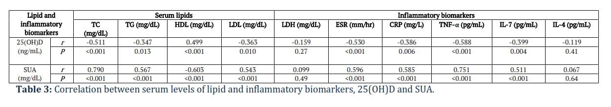
We found that serum level of 25(OH)D was inversely correlated with TC (P<0.001), TG (P=0.013), LDL (P=0.010), ESR (P<0.001), CRP (P=0.006), TNF-α (P<0.001), IL-7 (P=0.004) and was positively correlated with HDL (P<0.001), Table 3. Moreover, SUA was positively correlated with TC, TG, LDL, ESR, CRP, TNF-α, IL-7 (P<0.001, each) and was inversely correlated with HDL (P<0.001) existed, Table 3.
Regarding WOMAC scores, patients with grades II and III Kallgren scores had significantly higher WOMAC scores than grade I patients (39.73±7.78, 69.75±15.18 vs 18.11±7.22, respectively, P<0.001), Figure 1a. Additionally, WOMAC scores were inverse correlated with serum (25(OH)D) levels (P<0.001), Figure 1b. In contrast, WOMAC scores were positively correlated with serum SUA levels (P<0.001), Figure 1c.
The ROC curve analysis revealed that serum (25(OH)D) levels can excellently differentiate between moderate-to-severe (grades II and III) KOA women and those with mild KOA (grade I), an AUC of 0.955. [(95% confidence interval (CI): 0.872 to 1.0, P<0.001)]. The optimal sensitivity and specificity were (96.77% and 94.74%, respectively at a cutoff expression value<26.5 ng/mL), Figure 1d. Similarly, SUA can differentiate between moderate-to-severe (grades II and III) KOA women and those with mild KOA (grade I), with an AUC of 0.760, [(95% (CI): 0.620 to 0.899, P=0.002), an accepted discrimination ability. The optimal sensitivity and specificity were (77.42% and 73.68%, respectively at a cutoff expression value>8.67 mg/dL), Figure 1e.
Figures & Tables


Our study proved significant associations between serum levels of 25(OH)D and SUA with serum levels of lipids and inflammatory cytokines in obese elderly women with KOA. In addition, serum levels of 25(OH)D and uric acid can serve as reliable simple reliable prognostic tools in the prediction of KOA severity based on Kellgren scores in those women. However, serum 25(OH)D revealed better prognostic performance compared with SUA.
Synovitis, associated with OA, is incriminated in the severity and progression of pain and structural damage of OA. Moreover, increased inflammatory/immune cells including macrophages and lymphocytes have been evidenced in OA synovium [29].
Hyperuricemia is an inflammatory condition that can cause deleterious health effects such as gout and cardiovascular diseases. High SUA levels can cause joint inflammation and pain secondary to the formation of crystals [30]. Vitamin D deficiency is prevalent, especially among the elderly. The body’s inflammatory responses are crucial in developing and advancing KOA. It is believed that insufficient vitamin D levels may contribute to the pathogenesis of OA [11]. Moreover, vitamin D deficiency can induce parathyroid hormone release leading to increase SUA [31].
Functions of vitamin D includes maintaining normal bone homeostasis and enhancing the intestinal absorption of calcium, phosphorous, magnesium, iron, and zinc. In addition, vitamin D has several physiological functions such as control of insulin and glucose secretion and regulation of cardiorenal functions. Moreover, it has anti-inflammatory and immunomodulatory functions [32–36]. In corroboration to the current results, several epidemiological studies have revealed a positive association between vitamin D deficiency and chronic inflammatory disorders, e.g., chronic renal conditions, hepatic inflammatory diseases, inflammatory bowel diseases and rheumatoid arthritis [29,37].
Our findings revealed that CRP and ESR levels are helpful indicators of inflammation and tissue damage in KOA rather than LDH. In addition, they showed higher levels among severe than mild cases. Many studies agree with this observation [38,39].
Coinciding with our findings, Amirkhizi and colleagues [40] in their previous work reported similar findings. We reported that patients with grades II and III Kallgren scores had significantly lower serum 25(OH)D levels than grade I patients. In support, Heidari et al., [41] clarified that the severity of OA is positively associated with vitamin D level. A meta-analysis, conducted by Yu et al., [11] concluded similar results.
Furthermore, the current findings showed that vitamin D levels are inversely correlated to CRP and ESR levels in KOA patients. In support, vitamin D deficiency is accompanied by overexpression of the inflammatory markers [42]. The significant positive correlations between SUA levels and CRP and ESR implying that elevated SUA levels may be associated with increased systemic inflammation. Activation of the proinflammatory NF-κB signalling cascade could be induced by hyperuricemia [43]. We found a significant negative correlation between serum 25(OH)D and functional disability in KOA patients existed. This agreed with the previous finding of saturation vitamin D stores may relieve joint pain in case of vitamin D deficiency [44].
Moreover, serum UA >8.67 mg/dL can differentiate moderate-severe KOA from mild cases with less sensitivity and specificity than 25(OH)D (77.42% and 73.68% respectively). An American study of KOA subjects with asymptomatic hyperuricemia showed that SUA levels significantly distinguished patients with disease progression from non-progressors based on radiography over one year period of follow-up [45]. The association of SUA and OA has long been evaluated, and the pathological similarity between gout and OA has been proposed. Aging and obesity are significant predictors shared by both diseases [46–48]. According to Ding and coworkers [49] elevated levels of SUA were positively associated with the most radiographic alterations with KOA in both males and females patients including osteophytes but not joint space narrowing. In contrast to our results, in Korean population-based epidemiological study, SUA was not considered as a significant determinant for KOA progression [50].
According to our findings, vitamin D levels are linked to serum lipid components. Vitamin D positively correlates with serum HDL, whereas TC, TG, and LDL correlate negatively. Several studies supported this result and stated that less vitamin D levels are associated with an atherogenic lipid profile [51,52]. Multiple cardiovascular and metabolic factors can increase the risk of OA, causing metabolic syndrome. This syndrome includes vascular alteration, decreased HDL levels, increased both LDL and TG levels, elevated blood glucose levels and abdominal obesity, creating a low-grade chronic inflammatory condition [53–55]. Raised SUA levels are commonly observed in patients with metabolic syndrome. A former meta-analysis including 32 studies found a bidirectional association between SUA and vitamin D [56,57]. Patients with early OA experienced higher levels of LDL, UA, and CRP and significantly correlated with higher levels of pain intensity and higher disability [58].
An open-label clinical trial enrolled 80 patients with KOA by Divjack et al., [59] demonstrated improved clinical response as assessed by the visual analogue scale pain and WOMAC scores three months following vitamin D administration in those patients. A 3-month oral vitamin D supplementation was associated with decreased serum levels of inflammatory cytokines as TNF-α. UA could stimulate an innate immune response contributing to OA pathology and progression. A randomized controlled trial of Leung and colleagues [60] concluded that low dose colchicine reduced inflammation and high bone turnover but did not cause clinical improvement in most patients.
Overall, these results suggested that metabolic and inflammatory markers are chief players in the pathogenesis of KOA and act as potential targets for novel treatment measures. The findings also indicated the need for early detection and management of serum metabolic and inflammatory changes in patients with KOA to improve outcomes and quality of life as increased UA levels and/or reduced vitamin D levels are linked to worsened functional impairment with KOA.
Our study had some limitations. First, all the enrolled women were obese. Therefore, some laboratory findings might be confounded by the effect of obesity. Second, the lack of the follow-up assessments of the relevant biomarkers and the functional status of KOA patients confers another limitation.
Our results suggest robust associations between vitamin D deficiencies, high SUA levels with increased serum lipids and proinflammatory biomarkers as well as increased progression of clinical symptoms and radiological findings in KOA obese elderly women. In addition, serum levels of 25(OH)D and uric acid can serve as promising simple reliable prognostic tools in the prediction of KOA severity based on radiographic grading in those women. However, the prognostic performance of serum 25(OH)D was superior to SUA.
Ethical Statement
The Scientific Research Ethics Committee at King Faisal Medical Complex approved the study proposal (Ethical Permission No. 2023-B-1). Informed written consent was obtained from all participants. The study was following the 1964 Helsinki Declaration and its later amendments.
Author Contributions
AFG conceived the study, study design, participated in data collection, blood samples collection, performing the biochemical analyses. The initial draft was written by AFG, EIA, and ON performed data analysis and wrote the original draft. AAA, RAA, NAM, AA, AA, OA, MA, and AFH shared in blood samples collection, performing the biochemical analyses, AHA participated in data collection. All authors contributed to drafting, and reviewing, and approved the final version.
The author declare that there is no conflict of interest regarding the publication of this paper.
![]() References
References
- Palazzo C, Nguyen C, Lefevre-Colau MM, Rannou F, Poiraudeau S. Risk factors and burden of osteoarthritis. Annals of Physical and Rehabilitation Medicine, (2016); 1;59(3):134-138.
- Rahmati M, Nalesso G, Mobasheri A, Mozafari M. Aging and osteoarthritis: central role of the extracellular matrix. Ageing Research Reviews, (2017)1;40:20-30.
- Loeser RF, Collins JA, Diekman BO. Ageing and the pathogenesis of osteoarthritis. Nature Reviews Rheumatology, (2016);12(7):412-420.
- Puig-Junoy J, Zamora AR. Socio-economic costs of osteoarthritis: a systematic review of cost-of-illness studies. Seminars in Arthritis and Rheumatism, (2015); 44(5): 531-541.
- Liu S, Deng Z, Chen K, Jian S, Zhou F, et al. Cartilage tissue engineering: from proinflammatory and anti inflammatory cytokines to osteoarthritis treatments. Molecular Medicine Reports, (2022); 25(3):99.
- Puenpatom RA, Victor TW. Increased prevalence of metabolic syndrome in individuals with osteoarthritis: an analysis of NHANES III data. Postgraduate medicine, (2009)1;121(6):9-20.
- Mabey T, Honsawek S. Role of vitamin D in osteoarthritis: molecular, cellular, and clinical perspectives. International Journal of Endocrinology, (2015): 383918.
- Wicherts IS, Van Schoor NM, Boeke AJ, Visser M, Deeg DJ, et al. Vitamin D status predicts physical performance and its decline in older persons. The Journal of Clinical Endocrinology & Metabolism (2007)1;92(6):2058–2065.
- Sanghi D, Mishra A, Sharma AC, Singh A, Natu SM, et al. Does vitamin D improve osteoarthritis of the knee: a randomized controlled pilot trial. Clinical Orthopaedics and Related Research, (2013);471(11):3556–3562.
- Cao Y, Winzenberg T, Nguo K, Lin J, Jones G, et al. Association between serum levels of 25-hydroxyvitamin D and osteoarthritis: a systematic review. Rheumatology, (2013);52(7):1323–1334.
- Yu Y, Liu D, Feng D, Zhao J. Association between vitamin D and knee osteoarthritis: A PRISMA-compliant meta-analysis. Zeitschrift fur Orthopadie und Unfallchirurgie, (2021);159(3):281–287.
- Abbas MA. Physiological functions of Vitamin D in adipose tissue. The Journal of Steroid Biochemistry and Molecular Biology, (2017); 165(Pt B), 369–381.
- Zhu Y, Pandya BJ, Choi HK. Prevalence of gout and hyperuricemia in the US general population: the National Health and Nutrition Examination Survey 2007-2008. Arthritis and Rheumatism, (2011);63(10):3136–3141.
- Qi D, Liu J, Wang C, Wang L, Zhang X, et al. Sex-specific differences in the prevalence of and risk factors for hyperuricemia among a low-income population in China: A cross-sectional study. Postgraduate Medicine, (2020);132(6), 559–567.
- Lu J, Sun M, Wu X, Yuan X, Liu Z, et al. Urate‐lowering therapy alleviates atherosclerosis inflammatory response factors and neointimal lesions in a mouse model of induced carotid atherosclerosis. The FEBS Journal, (2019);286(7):1346–1359.
- Boehme KA, Rolauffs B. Onset and Progression of human osteoarthritis—can growth factors, inflammatory cytokines, or differential mirna expression concomitantly induce proliferation, ECM degradation, and inflammation in articular cartilage?, International Journal of Molecular Sciences, (2018);19(8):2282.
- Sirikaew N, Chomdej S, Tangyuenyong S, Tangjitjaroen W, Somgird C, et al. Proinflammatory cytokines and lipopolysaccharides up regulate MMP-3 and MMP-13 production in Asian elephant (Elephas maximus) chondrocytes: attenuation by Anti-arthritic agents. BMC Veterinary Research, (2019);15(1):419.
- Wang T, He C. Pro-inflammatory cytokines: The link between obesity and osteoarthritis. Cytokine Growth Factor Reviews, (2018);44:38–50.
- Nguyen L, Sharma A, Chakraborty C, Saibaba B, Ahn ME, et al. Review of prospects of biological fluid biomarkers in osteoarthritis. International Journal of Molecular Sciences, (2017);18(3):601.
- Mabey T, Honsawek S, Tanavalee A, Yuktanandana P, Wilairatana V, et al. Plasma and synovial fluid inflammatory cytokine profiles in primary knee osteoarthritis. Biomarkers, (2016);21(7):639–644.
- Liu S, Cao C, Zhang Y, Liu G, Ren W, et al. PI3K/Akt inhibitor partly decreases TNF-α-induced activation of fibroblast-like synoviocytes in osteoarthritis. Journal of Orthopaedic Surgery and Research, (2019);14(1):425.
- Abdel-Naby HM, El-Tawab SS, Rizk MM, Aboeladl NA. Is interleukin-17 implicated in early knee osteoarthritis pathogenesis as in rheumatoid arthritis? Egyptian Rheumatology and Rehabilitation, (2022);49(1):29.
- Schuerwegh A, Dombrecht E, Stevens W, Van Offel J, Bridts C, et al. Influence of pro-inflammatory (IL-1α, IL-6, TNF-α, IFN-γ) and anti-inflammatory (IL-4) cytokines on chondrocyte function. Osteoarthritis and cartilage, (2003);11(9):681–687.
- Wojdasiewicz P, Poniatowski ŁA, Szukiewicz D. The role of inflammatory and anti-inflammatory cytokines in the pathogenesis of osteoarthritis. Mediators of Inflammation, (2014); 2014(1):561459.
- Altman R, Asch E, Bloch D, Bole G, Borenstein D, et al. Development of criteria for the classification and reporting of osteoarthritis: Classification of osteoarthritis of the knee. Arthritis & Rheumatology, (1986);29(8):1039–1049.
- Alghadir A, Anwer S, Iqbal ZA, Alsanawi HA. Cross-cultural adaptation, reliability and validity of the Arabic version of the reduced Western Ontario and McMaster Universities Osteoarthritis index in patients with knee osteoarthritis. Disability and Rehabilitation, (2016);38(7):689–694.
- Kellgren JH, Lawrence JS. Radiological assessment of osteo-arthrosis. Annals of the Rheumatic Diseases, (1957); 16(4):494–502.
- Gilmour D, Sykes AJ. Westergren and Wintrobe methods of estimating ESR compared. British Medical Journal, (1951);2(4746):1496–1497.
- Park CY. Vitamin D in the prevention and treatment of osteoarthritis: from clinical interventions to cellular evidence. Nutrients, (2019);11(2):243.
- Shahin L, Patel KM, Heydari MK, Kesselman MM. Hyperuricemia and cardiovascular risk. Cureus, (2021);13(5): e14855.
- Zhang YY, Qiu HB, Tian JW. Association between vitamin D and hyperuricemia among adults in the United States. Frontiers in Nutrition, (2020);7:592777.
- Liu PT, Stenger S, Li H, Wenzel L, Tan BH, et al. Toll-Like receptor triggering of a vitamin D-mediated human antimicrobial response. Science, (2006); 311(5768):1770–1773.
- Holick MF. Vitamin D deficiency. New England Journal of Medicine, (2007);357(3):266–281.
- Marwaha RK, Tandon N, Reddy DRH, Aggarwal R, Singh R, et al. Vitamin D and bone mineral density status of healthy schoolchildren in northern India. The American journal of Clinical Nutrition, (2005);82(2):477–482.
- Garbossa SG, Folli F. Vitamin D, sub-inflammation and insulin resistance. A window on a potential role for the interaction between bone and glucose metabolism. Reviews in Endocrine and Metabolic Disorders, (2017);18(2):243–258.
- Maestro B, Campión J, Dávila N, Calle C. Stimulation by 1,25-dihydroxyvitamin D3 of insulin receptor expression and insulin responsiveness for glucose transport in U-937 human promonocytic cells. Endocrine Journal, (2000);47(4):383–391.
- Vos T, Flaxman AD, Naghavi M, Lozano R, Michaud C, et al. Years lived with disability (YLDs) for 1160 sequelae of 289 diseases and injuries 1990–2010: a systematic analysis for the Global Burden of Disease Study 2010. The lancet, (2012);380(9859):2163-2196.
- Hanada M, Takahashi M, Furuhashi H, Koyama H, Matsuyama Y. Elevated erythrocyte sedimentation rate and high-sensitivity C-reactive protein in osteoarthritis of the knee: relationship with clinical findings and radiographic severity. Annals of Clinical Biochemistry, (2016); 53(5):548-553.
- Jin X, Beguerie J, Zhang W, Blizzard L, Otahal P, et al. Circulating C-reactive protein in osteoarthritis: a systematic review and meta-analysis. Osteoarthritis and Cartilage, (2014);22:S293.
- Amirkhizi F, Asoudeh F, Hamedi-Shahraki S, Asghari S. Vitamin D status is associated with inflammatory biomarkers and clinical symptoms in patients with knee osteoarthritis. Knee, (2022);36:44–52.
- Heidari B, Heidari P, Hajian-Tilaki K. Association between serum vitamin D deficiency and knee osteoarthritis. International orthopaedics, (2011);35(11):1627–1631.
- Etminan A, Seyed Askari SM, Naghibzade Tahami A, Adel Mahdi S, Behzadi M, et al. Relationship between the serum levels of Vitamin D and inflammatory markers in ESRD patients. Acta Biomedica: Atenei, (2020);91(4):e2020099.
- Spiga R, Marini MA, Mancuso E, Di Fatta C, Fuoco A, et al. Uric acid is associated with inflammatory biomarkers and induces inflammation via activating the NF-κB signaling pathway in HepG2 cells. Arteriosclerosis, Thrombosis, and Vascular Biology. (2017);37(6):1241-1249.
- Laslett LL, Quinn S, Burgess JR, Parameswaran V, Winzenberg TM, et al. Moderate vitamin D deficiency is associated with changes in knee and hip pain in older adults: a 5-year longitudinal study. Annals of the Rheumatic Diseases, (2014);73(4):697-703.
- Krasnokutsky S, Oshinsky C, Attur M, Ma S, Zhou H, et al. Serum urate levels predict joint space narrowing in non‐gout patients with medial knee osteoarthritis. Arthritis & Rheumatology, (2017);(6):1213-1220.
- Roddy E, Doherty M. Gout and osteoarthritis: a pathogenetic link? Joint Bone Spine, (2012);79(5), 425–427.
- Blagojevic M, Jinks C, Jeffery A, Jordan KP. Risk factors for onset of osteoarthritis of the knee in older adults: a systematic review and meta-analysis. Osteoarthritis and Cartilage, (2010);18(1):24–33.
- Choi HK, Atkinson K, Karlson EW, Curhan G. Obesity, weight change, hypertension, diuretic use, and risk of gout in men. . Archives of Internal Medicine, (2005);165(7), 742–748.
- Ding X, Zeng C, Wei J, Li H, Yang T, et al. The associations of serum uric acid level and hyperuricemia with knee osteoarthritis. Rheumatolology International, (2016);36(4):567–673.
- Go DJ, Kim DH, Kim JY, Guermazi A, Crema MD, et al. Serum uric acid and knee osteoarthritis in community residents without gout: A longitudinal study. Rheumatology, (2021);60(10):4581–4590.
- Lupton JR, Faridi KF, Martin SS, Sharma S, Kulkarni K, et al. Deficient serum 25-hydroxyvitamin D is associated with an atherogenic lipid profile: The Very Large Database of Lipids (VLDL-3) study. Journal of Clinical Lipidology, (2016);10(1):72-81.
- Asbaghi O, Kashkooli S, Choghakhori R, Hasanvand A, Abbasnezhad A. Effect of calcium and vitamin D co-supplementation on lipid profile of overweight/obese subjects: a systematic review and meta-analysis of the randomized clinical trials. Obesity Medicine, (2019);15:100124.
- Bierma-Zeinstra SM, Koes BW. Risk factors and prognostic factors of hip and knee osteoarthritis. Nature Clinical Practice. Rheumatology, (2007);3(2),78–85.
- Luppino FS, de Wit LM, Bouvy PF, Stijnen T, Cuijpers P, et al. Overweight, obesity, and depression. Archives of General Psychiatry, (2010);67(3), 220–229.
- Festa A, D’Agostino R, Howard G, Mykkänen L, Tracy RP, et al. Chronic subclinical inflammation as part of the insulin resistance syndrome. Circulation, (2000);102(1):42–47.
- Copur S, Demiray A, Kanbay M. Uric acid in metabolic syndrome: Does uric acid have a definitive role? European Journal of Internal Medicine, (2022);103:4–12.
- Isnuwardana R, Bijukchhe S, Thadanipon K, Ingsathit A, Thakkinstian A. Association between vitamin D and uric acid in adults: a systematic review and meta-analysis. Hormone and Metabolic Research, (2020);52(10):732–741.
- Herrero-Manley L, Alabajos-Cea A, Suso-Martí L, Cuenca-Martínez F, Calatayud J, et al. Serum lipid biomarkers and inflammatory cytokines associated with onset and clinical status of patients with early knee osteoarthritis. Frontiers in Nutrition, (2023),10, 1126796.
- Divjak A, Jovanovic I, Matic A, Lucic AT, Gajovic N, et al. The influence of vitamin D supplementation on the expression of mediators of inflammation in knee osteoarthritis. Immunologic Research, (2023);71(3):442–450.
- Leung YY, Haaland B, Huebner JL, Wong SB, Tjai M, et al. Colchicine lack of effectiveness in symptom and inflammation modification in knee osteoarthritis (COLKOA): a randomized controlled trial. Osteoarthritis Cartilage, (2018);26(5):631–640.
This work is licensed under a Creative Commons Attribution-Non Commercial 4.0 International License. To read the copy of this license please visit: https://creativecommons.org/licenses/by-nc/4.0

