

Full Length Research Article
Exploring Listeria monocytogenes in Ewe Milk: ssrA Gene-based Real-time PCR Identification, Phylogenetic Analysis, and Antibacterial Assessment of Magnesium Oxide Nanoparticles Synthesized with Myrtus communis Leaf Extract
Jenan Nadhim Sadeq, Balsam Miri Mizher, Alyaa Abdulhussein Alsaedi*, Ola Hakim Khudhair
Adv. life sci., vol. 11, no. 3, pp. 634-640, August 2024
*- Corresponding Author: Alyaa Abdulhussein Alsaedi (Alyaa.alsaedi@qu.edu.iq)
Authors' Affiliations
[Date Received: 16/11/2023; Date Revised: 29/06/2024; Date Published: 10/07/2024]
Abstract![]()
Introduction
Methods
Results
Discussion
References
Abstract
Background: Listeria monocytogenes, a zoonotic pathogen affecting humans and animals, exhibits a global distribution, including Iraq. This study focused on the rapid identification and phylogenetic analysis of L. monocytogenes in freshly collected ewe milk samples from various farms in Al-Qadisiyah province, Iraq.
Methods: The study was conducted with care and precision, involving 150 milk samples. These samples were subjected to traditional bacterial isolation and identification using the enrichment culture method and biochemical tests, with the PCR technique confirming the results. The antibacterial activity of MgONPs was then assessed using the disc diffusion method, ensuring a comprehensive and reliable approach to the study.
Result: The results show that 150 ewe milk samples underwent real-time PCR (RT-PCR) targeting the ssrA gene, followed by partial 16S rRNA gene sequencing (PSGS) of purified conventional PCR products. Furthermore, the study entails the biosynthesis of magnesium oxide nanoparticles using Myrtus communis leaf extract, followed by a comprehensive characterization utilizing UV-spectra, FTIR, SEM, and TEM techniques. The Agar well diffusion method assessed the antibacterial efficacy of these Biosynthesized nanoparticles against L. monocytogenes. The RT-PCR results revealed the presence of L. monocytogenes in 36 out of 150 samples (24%). Subsequent PCR analysis confirmed the presence of the pathogen in 30 out of these 36 positive samples (83.33%). Sequencing of two purified PCR products demonstrated 100% nucleotide identity with global isolates from Iraq and Turkey. Furthermore, the study demonstrated that L. monocytogenes exhibited substantial sensitivity (24.66 ± 0.3) to the biosynthesized magnesium oxide nanoparticles. These findings underscore the speed and precision of the RT-PCR method for detecting L. monocytogenes in fresh ewe milk samples.
Conclusion: This comprehensive investigation enhances our understanding of L. monocytogenes prevalence in ewe milk and highlights the potential of Myrtus communis -derived nanoparticles for combating this pathogen.
Keywords: Antibacterial nanoparticles; Listeria monocytogenes; Magnesium oxide; Myrtus communis sheep milk; ssrA gene
Introduction![]()
L. monocytogenes is the listeriosis-etiological agent, a serious infection with substantial hospitalization and death rates in both humans and animals. Mainly L. ivanovii and L. monocytogenes are pathogenic bacteria of mammals, however, there are 21 species of these little rod-shaped gram-positive bacterial microorganisms in the genus Listeria. Outbreaks of human and animal listeriosis have had huge financial consequences on the social system and the food business since the pathogenic species evolved as a prominent foodborne pathogen in Western nations. Listeria-based infections have risen in Europe since 2008, initiating a major public health concern [1,2].
Even though the Listeria genus originated as saprophytic bacteria, certain species have effectively adjusted to other ecological niches linked with human practices, such as farms, food, and food-processing settings. Listeria may infect mammalian hosts through contaminated food. L. monocytogenes, once inside the host, uses a wide variety of complex processes to penetrate and live within eukaryotic cells, avoid being destroyed by the host's immune system, and eventually spread throughout the body. This bacterium may pass to the brain and placenta, leading to meningitis or abortion [3-6]. Moreover, like many other microorganisms, evidence of antibacterial resistance in L. monocytogenes isolates from food sources is reported, which poses a serious threat to future treatment of listeriosis [7].
Antibiotic resistance is a critical issue that has necessitated the use of drugs that are progressively more expensive, toxic, and ineffective. Several mechanisms can contribute to antibiotic resistance of bacteria, involving a reduction in the drug uptake, modifications of the antibacterial target, activation of efflux mechanisms for extrusion of the antibacterial agents, and global alterations in prominent metabolic pathways. These scientists are turning to innovative solutions to overcome the antibiotic resistance problem. To overcome antibiotic resistance, Nanoparticles have been developed as novel that can utilized either directly or indirectly [8]. In this regard, nanobiotechnology is another area of research utilizing natural sources to make Nanoparticles (Nps) exhibit antibacterial potency [9]. Nanobiotechnology uses a biogenic reduction method in which a reducing agent is replaced by an extract of natural products with inherent stabilizing, growth terminating, and capping properties. Biosynthesis of NPs from metal ions is more environmentally friendly, free of chemical contamination, less expensive, and safe for biological applications [10]. Furthermore, compared to other biological processes, using plants for nanoparticle synthesis can be advantageous. It also eliminates the ease of large scale-up and the process of culture maintenance, and there is no need to use high pressure, energy, temperature, or toxic chemicals [11]. Nevertheless, some critical factors may affect the final accuracy of a PCR technique, such as the targeted genes, methods of DNA extraction, and specificity of primers. Moreover, a PCR test is only good for evaluating whether bacteria are present, and not for counting how many bacteria are there. The herein study was conducted to generate a fast RT-PCR dependent identification method and a phylogenetic study of L. monocytogenes from highly fresh field-collected ewe milk utilizing the ssrA gene as a molecular target.
Methods![]()
Sample collection
The present study recruited 150 raw ewe milk samples from different farms in Al-Qadisiyah Province. These samples were collected under sterile conditions, including wash-cleaning and 70%-ethanol-disinfecting the udder teats. The samples were placed in 25ml sterile containers and sent to the Laboratory of Microbiology, College of Veterinary Medicine, University of Al-Qadisiyah, Al-Diwaniyah City, Iraq.
Isolation and identification L. monocytogenes
Out of 150 milk samples, 36 tested positives for L. monocytogenes using real-time PCR. These 36 PCR-positive milk samples were selected for further analysis. Each sample was streaked onto Modified Oxford agar (MOX) medium and incubated at 35°C for 24 hours. After the incubation period, the colony morphology of the isolates was carefully observed. The identification of isolates was confirmed through microscopic examination of Gram-stained smears and a series of biochemical tests. These tests included hemolysis on blood agar, indole test, enzyme assays (catalase, urease, oxidase), aesculin hydrolysis test, and sugar fermentation tests (lactose, glucose, D-xylose, L-rhamnose, mannitol, arabinose). To further confirm the presence of L. monocytogenes, DNA extraction was performed from each isolate followed by PCR amplification and detection of the 16S rRNA gene. Additionally, amplified DNA fragments were sequenced to ensure accurate species identification.
Extraction of Listeria monocytogenes genomic DNA
Bacterial genomic DNA was extracted from ewe milk directly by using the PrestoTM Mini gDNA Bacteria Kit (Geneaid, USA). The supernatant resulted from a centrifugation process at 10000 rpm for 2 mins of 1 ml per milk sample was discarded. The bottom fat layer was used for the DNA extraction based on the kit manufacturer's requirements. The DNA was estimated for purity and amount using a Nanodrop. The DNA was then kept at -20˚C until conducting the RT-PCR technique.
Real-time PCR
A Syber green dye-based RT-PCR amplification was performed employing the ssrA gene as a target at 130bp. The primers (GenBank: CP054040.1) were purchased from Bioneer Company (Korea). The sequence of these primers was F: ACCCTTACCGTAGCACATGG and R: GGGATCGTCCTCGTTATCAA. The reaction of RT-PCR was done by utilizing the AccuPowerTM 2X kit (Bioneer company, Korea) and based on its accompanying protocol, which included 50µl total volume (2.5µl bacterial DNA, 25µl (2X) green star master mix, 2µl (10pmol)/each primer direction, and 18.5µl water for molecular use. These components were placed in sterile tubes and vortexed for 3 minutes. Then, the tubes were placed in a MiniOpticon RT-PCR system using the following thermocycler conditions: (95˚C-180s)-one-cycle initial denaturation, 40 cycles of
(95˚C-10s)-denaturation and (58˚C-30s)-annealing/extension/detection, and (65˚C-95˚C-0.5s)-one cycle melting.
Conventional PCR for gene sequencing
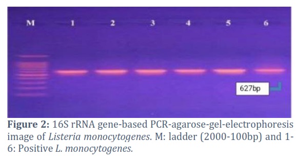
The primers (F: GCCAGCCAAGGAGCATTAT and R: GCCCAGTTTAACGCATCTTC) that target a 16S rRNA genetic piece at 627bp were used. The PCR products were visualized under UV light.
Listeria monocytogenes-based 16S rRNA gene sequencing
Two 16S rRNA-PCR products were sequenced at the Bioneer Company (Korea) after being extracted from the agarose gel using the EZ EZ-10 Spin Column DNA Gel Extraction Kit (Biobase, Canada). These sequences were submitted to the NCBI, aligned by (BLAST), and the genetic tree analysis and tree were performed using MEGA software.
Biosynthesis of nanoparticles
The "bottom-up" approach for nanoparticles in the synthesis Biosynthesis of MgONPs using plant extracts (Myrtus communis ). The bottom-up approach involves building nanoparticles from smaller components or atoms, gradually assembling them into the desired nanoscale structure. In the case of green synthesis, the process begins with bioactive compounds present in the plant extract, which act as reducing agents to reduce metal ions into nanoparticles. The nanoparticles are essentially "grown" from these smaller components, building up their structure through the reduction and stabilization processes provided by the plant extract [13].
Preparation of the extract
Myrtus communis leaves were collected from the local gardens of al Diwaniyah Province, Iraq in May/ 2022. Leaves were shade-dried for a week and then ground to fine powder weighing about 20 gm. After 30 min of boiling in 100 ml of distilled water in a round bottom flask, the powdered leaves were allowed to be cooled at room temperature. Then, Whatman filter paper was used to filter the resultant mixture in order to purify the extract. The filtrate was stored under 4°C for later investigations.
Synthesis of MgONPs
MgOPs were synthesized thus; In a conical flask 50 ml of 1M of Mg (NO3)2 slowly added in magnetic stirring to 50ml of leaf solution at 80°C for 4 hrs till the solution converted to brownish colloidal color. The test is the time for complete conversion of MgONP’s brownish brown color is 2 hrs to ( 20%), 2h 30min to ( 79 %) and 4h to 100 %. The subject material was then centrifuged for magnetization at the speed of 15,000 rpm in 10 Min for separate out OM; the subject material was then dried in the oven at 50 °C. After the drying calcination is done in 400 °C in muffle furnace for conversion to the powder white colored MgONPs. FTIR, SEM, TEM, UV–spectra, and other various spectral analyses believe the structure of MgOPs. The UV–vis absorbance spectrum of MgONPs was obtained by UV–vis spectrophotometer, device with prominent characters, Shimadzu UV–vis spectrophotometer, (UV-1800, Japan), had measure resolution 1 nm, Wavelength range (200–800 nm) was attained by the measuring procedure. The dimensions of the MgONPs were carried out by transmission electron microscopy (TEM) using (TECNAI G-20 instrument), Scanning Electron Microscopy (SEM) by instrument (Nova nano FE-SEM 450 FEI). The degree of purification of Myrtus communis extract was investigated by the removal of impurities by this UV–spectra presented. In FT-IR by using an FT-IR instrument (PerkinElmer Spectrum 2000 FTIR; KBr pellet technique, resolution 2 cm, range 4000–400 cm−1).
Antibacterial activity of MgONPs
The results on antibacterial activity of MgONPs were performed by disc diffusion method. The bacterial test microflora, cultivated and found in nutrient broth medium, was utilized to perform further experiments in the nutrient broth medium. The nutrient agar plates were prepared, sterilized and solidified. Then, 1 ml of Listeria monocytogenes bacteria (1.3 × 10⁵ colony-forming units (CFU/ml) were spread on the prepared Petri plates by sterile glass rod having aim to developed bacterial colony. Each plate was prepared with four wells (6 mm diameter) using corked borer. An equal amount of 10µl MgONPs, 10µl of MgNO3, 10µl Plant extract only, and 10µl distill water were added in each well. Plates were incubated for 24 h at 37 °C and an inhibition zone was observed around the wells. After the test were conducted three times.
Results![]()
Isolation and identification
From the total tested milk samples, 30 samples (83.3%) had a well-grown MOX agar. Diagnostic microbiological tests were performed for 30 samples with an initial positive culture, and all isolates were identified as L. monocytogenes based on Gram-positive bacillus. On the other hand, the results of biochemical tests showed a positive reading for each esculin hydrolysis, β-hemolysis, catalase, and acid production from [L. rhamnosus, lactose, glucose] while the recorded negative result for fermentation (D-xylose, mannitol and arabinose), oxidase, urease and Indole test [12].
RT-PCR
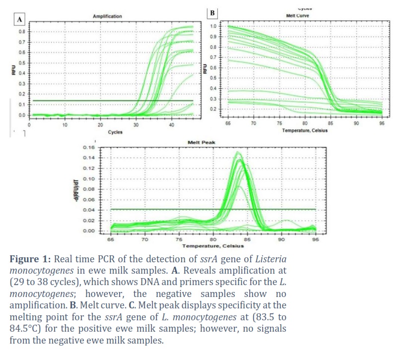
The herein study was conducted to generate a fast RT-PCR-dependent identification method of L. monocytogenes from highly fresh field-collected ewe milk utilizing the ssrA gene as a molecular target. The findings of the RT-PCR revealed 36/150 (24%) positive samples regarding the presence of L. monocytogenes. The results of the RT-PCR showed that the time required for the complete run to identify L. monocytogenes was significantly (p˂0.05) reduced to half. The findings of the RT-PCR for the detection of ssrA gene of Listeria monocytogenes in ewe milk samples revealed amplification at (29 into 38 cycles), which showed that the DNA template and primers were specific for the L. monocytogenes; however, the negative samples showed no amplification (Figure 1A). Moreover, the melt curve and melt peak recorded specificity at the melting point for the ssrA gene of L. monocytogenes at (83.5 to 84.5°C) for the positive ewe milk samples; however, no signals were revealed from the negative ewe milk samples (Figure 1A) and Figure 1B). Significant (p˂0.0001) sensitivity and specificity were detected for the current RT-PCR method for the L. monocytogenes identification in the examined ewe milk samples. The PCR recorded 30/36 (83.33%) positive samples for the occurrence of the pathogen (Figure 2).
Phylogenetic Analysis
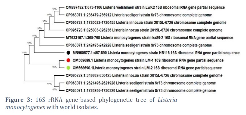
16S rRNA gene-based phylogenetic tree of Listeria monocytogenes with world isolates has shown two isolates OM568689.1 and OM568690.1 are at 96% while one isolates MN960577.1 has shown 97% proximity in the tree (Figure 3). The phylogenetic tree suggests a relatively close genetic relationship between these two isolates. This proximity may indicate a recent common ancestor or shared evolutionary history. While the difference in proximity might seem small, it could have important implications for understanding the epidemiology and virulence of these strains.
UV–Vis Spectroscopy
The colloidal stability of the biosynthesized MgONPs was valuated and their validity was confirmed using UV–visible (UV–vis) spectroscopy. The resultant white solid powder was subjected to UV–vis analysis, yielding an absorption peak at 320 nm (Figure 4B). This specific wavelength is indicative of the presence of relatively small-sized nanoparticles with good colloidal stability.
FT-IR Analysis
Fourier Transform Infrared (FT-IR) spectroscopy was employed to discern the chemical composition of both Myrtus communis leaf extract and the synthesized magnesium oxide nanoparticles (MgONPs). At wavelengths ranging from 4000 to 400 cm-1 Spectral data were collected. The presence of hydroxyl functional groups that are inherent to alcohols and phenolic compounds. As indicated in strong absorption bands were identified at approximately 3417 cm-1. Thereafter, significant absorption bands appeared at wavenumbers of 3417, 2360, 1735, 1620, 1450, 1380, 1103, 1041, 609, 478, and 439 cm-1 in the FTIR spectrum of the MgONPs. These observations are suggestive of the participation of multiple functional groups derived from the Myrtus communis extract, acted as both reducing and stabilizing agents during the synthesis of the MgONPs. Plausible functional groups encompass esters, ethers, carbonyl (polyol derivatives), or aromatic constituents. The peak of absorption at 3417 cm-1 is ascribed to hydrogen-bonded hydroxyl and free hydroxyl groups, whereas the peak at 1620 cm-1 represents the stretching vibration of aromatic C=C bonds.
SEM and TEM Analysis
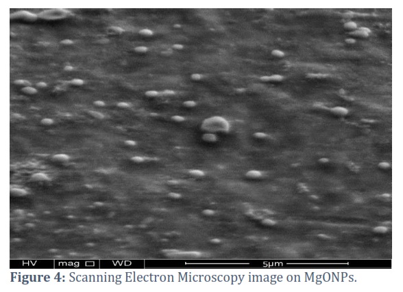
The morphology of the synthesized MgONPs was meticulously elucidated through scanning electron microscopy (SEM). SEM analysis underscored the even distribution of MgONPs on the substrates, characterizing them as spherical and irregular entities that collectively showed a granular surface topology. Subsequently, transmission electron microscopy (TEM) was employed to glean further insights into the shape and morphology of the synthesized MgONPs. TEM imaging corroborated predominantly spherical MgONPs characterized by variable diameters, as shown in (Figure 4).
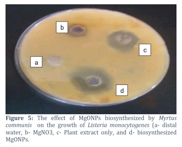
Antibacterial Activity
MgONPs can interact with bacterial cell membranes, disrupt cellular functions, and induce oxidative stress, ultimately leading to bacterial cell death. In vitro assessment of the antibacterial activity of MgONPs against L. monocytogenes was carried out, yielding that the effect of MgONPs biosynthesized by Myrtus communis on the growth of L. monocytogenes shows the values 8, 12.33,and 24.66 for treatment of AgNo3, plant extract and MgONPs consequently. The inhibition zone values, quantified in millimeters (mm), revealed that MgONPs showed a more pronounced inhibitory effect as compared to MgNO3, a counterpart substance tested against the same bacterial strain. Concurrently, it merits mention that the Myrtus communis leaf extract also manifested antibacterial activity; however, its inhibitory efficacy was notably less pronounced in comparison to MgONPs. This shows that the biosynthesized magnesium nanoparticles possess antibacterial potency (Figure 5).
Figures & Tables
Previous studies have advocated the PCR approaches and other molecular strategies for the detection of L. monocytogenes due to their high sensitivity, specificity, and in terms of speed [13-14]. Nevertheless, some critical factors may affect the final accuracy of a PCR technique, such as the targeted genes, methods of DNA extraction, and specificity of primers. Moreover, a PCR test is only good for evaluating whether bacteria are present, and not for counting how many bacteria are there [15].
Two purified PCR products were sequenced and deposited in the GenBank under the accession numbers OM568690.1, and OM568689.1. These isolates showed (100%) similar-nucleotide identity to global isolates from Iraq and Turkey with accession numbers of MN960577.1, and MT633107.1, respectively. However, less similarities were recorded with isolates from the USA, Egypt, and Poland with the accession numbers CP095728.1 (L. innocua), OM897482.1 (L. welshimeri), and CP063071.1 (L. seeligeri), respectively, (Figure 4). Rapid detection of contaminated food is crucial to restrict the spread of this pathogenic bacterial species during a foodborne epidemic. One or more molecular procedures must be performed to genotype the concerned bacteria, but which techniques could be used rely on certain geographical circumstances [16-17]. Molecular serogrouping was used to identify isolates before the introduction of RT-PCR. The screening turnaround time was reduced in half by using an RT-PCR-based approach. All the isolates that may be linked to the epidemic could be identified and analyzed in as little as two to four hours using the RT-PCR approach [18].
Traditional methods of determining whether species of Listeria are present require a significant amount of time and effort (operation takes 4-5 days) and rely on enrichment and selective media. Melting-curve analysis, multi-locus sequence typing, genome-sequencing, pulse-field gel electrophoresis, and PCR-based methodologies are just some of the molecular methods used to identify and classify this bacterium. Diagnostic tests should be conducted on this species [19-24]. Ka clíková developed a PCR-based test that detects Listeria at 10° CFU per 25gm of food after three days. The overall viable count recorded by [22] was 1.35±2×10⁸ and 0.35±1.9×10⁸ in salads and broccoli, respectively.
Listeria monocytogenes is known to be a genetically diverse species based on previous studies. [25-26]. This diversity can be ascribed to several factors including the potential for horizontal gene transfer and the adaptation to various environments, such as food processing facilities and natural environments. The 16S rRNA gene-based phylogenetic analysis provides insights into the relatedness of these isolates within the species. These results provide a foundation for further research into the genomic characteristics and epidemiological significance of these isolates, which can contribute to understanding Listeria monocytogenes evolution and its implications for public health.
On the other hand, the absorption peak is notable for being different from a previous study that used UV-vis spectroscopy, which was performed on a distinct plant extract sample and showed an absorption maximum of 245 nm [27].
The presence of MgOMg bonds is inferred from the stretching vibration mode at 609 cm-1. Moreover, it is worth noting that preceding research has substantiated those wavenumbers spanning 600-850 cm-1 typically correspond to magnesium oxide bonds, with the range of 400-520 cm cm-1 serving as corroboration of MgONPs formation [28-29].
The antibiotic antibacterial finding shows that the Spherical nanoparticles are generally considered more biocompatible than other shapes, making them potentially suitable for biomedical and drug delivery Applications. Furthermore, a comparative assessment of the antibacterial outcomes of MgONPs and MgNO3 vis-à-vis negative controls (distilled water) revealed a palpable difference, with MgONPs producing substantial inhibition zones as opposed to the leaf extract only and with the controls. This study is like the reviewed research studies [30-33], where MgONPs, biosynthesized by leave, showed microbial inhibition against multidrug-resistant (MDR) bacteria
This study addressed the pressing issue of Listeria monocytogenes infections, a significant threat to both human and animal health, and explored the potential of green-synthesized Magnesium Oxide Nanoparticles (MgONPs) as a novel antibacterial agent. This research encompassed various aspects, including the molecular detection of L. monocytogenes, phylogenetic analysis, and the biosynthesis and characterization of MgONPs, followed by an assessment of their antibacterial activity. A rapid RT-PCR-based approach has been employed, targeting the ssrA gene to detect L. monocytogenes in ewe milk samples. The results demonstrated the specificity and sensitivity of this method, offering a valuable tool for the quick and reliable identification of L. monocytogenes in dairy products. The study also conducted a 16S rRNA gene-based phylogenetic analysis, revealing the genetic relatedness of L. monocytogenes isolates. The analysis indicated a close genetic relationship between certain isolates, shedding light on their evolutionary history and potential epidemiological significance.
For the biosynthesis of MgONPs study adopted a "bottom-up" approach, utilizing Myrtus communis plant extract as a natural reducing agent for the synthesis of MgONPs. Characterization techniques for MgONPs, including UV-vis spectroscopy, FT-IR analysis, SEM, and TEM, confirmed the successful synthesis of predominantly spherical MgONPs with variable diameters. The UV-vis spectra suggested the presence of small-sized nanoparticles with good colloidal stability, a promising sign for potential applications. In vitro, assessment of the antibacterial activity of MgONPs against L. monocytogenes demonstrated their pronounced inhibitory effect. MgONPs exhibited greater antibacterial potency compared to MgNO3 and the plant extract alone, highlighting their potential as effective antibacterial agents.
In conclusion, our study contributes valuable insights into the detection and genetic analysis of L. monocytogenes, as well as the synthesis and characterization of MgONPs for antibacterial applications. These findings open doors to further investigations and applications of MgONPs in addressing bacterial infections, potentially improving public health and food safety. Additionally, our research underscores the significance of harnessing natural resources and green nanobiotechnology to address pressing challenges in microbiology and nanomedicine.
Author Contributions
Jenan Sadeq: Analysis, Writing – original draft
Balsam Al Muhana: Data curation, Supervision
Alyaa Abdulhussein Alsaedi: Conceptualization, Writing – review & editing
Ola Hakim Khudhair: Investigation, Project administration
The author declare that there is no conflict of interest regarding the publication of this paper.
![]() References
References
- ECDC, EFSA. The European Union One Health 2019 Zoonoses Report. European Food Safety Authority Journal, (2019); 17(12): e05926.
- Quereda JJ, Leclercq A, Moura A, Vales G, Gómez-Martín Á, et al. Listeria valentina sp. nov., isolated from a water trough and the faeces of healthy sheep. International Journal of Systematic and Evolutionary Microbiology, (2020); 70(11): 5868–79.
- Charlier C, Disson O, Lecuit M. Maternal-neonatal listeriosis. Virulence, (2020); 11(1): 391–7.
- NicAogáin K, O’Byrne CP. The Role of Stress and Stress Adaptations in Determining the Fate of the Bacterial Pathogen Listeria monocytogenes in the Food Chain. Frontiers in Microbiology, (2016); 7(11): 1865.
- Radoshevich L, Cossart P. Listeria monocytogenes: towards a complete picture of its physiology and pathogenesis. Nature Reviews Microbiology, (2018); 16(1): 32–46.
- Schlech WF. Epidemiology and Clinical Manifestations of Listeria monocytogenes Infection. Microbiology Spectrum, (2019); 7(3): 1–12.
- Al-Nabulsi A, Al-Holy MA, Shahbaz HM, Alimat AN, Abu Ghoush MH, et al. Emergence of antibiotic resistance in Listeria monocytogenes isolated from food products: a comprehensive review. Comprehensive Reviews in Food Science and Food Safety, (2018); 17(5): 1277-92.
- Anand U, Carpena M, Kowalska-Góralska M, Garcia-Perez P, Sunita K, et al. Safer plant-based nanoparticles for combating antibiotic resistance in bacteria: A comprehensive review on its potential applications, recent advances, and future perspective. Science of The Total Environment, (2022); 821: 153472.
- Castillo-Henríquez L, Alfaro-Aguilar K, Ugalde-Álvarez J, Vega-Fernández L, Montes de Oca-Vásquez G, et al. Green synthesis of gold and silver nanoparticles from plant extracts and their possible applications as antimicrobial agents in the agricultural area. Nanomaterials, (2020); 10(9): 1763.
- Soni V, Raizada P, Singh P, Cuong HN, Rangabhashiyam S, et al. Sustainable and green trends in using plant extracts for the synthesis of biogenic metal nanoparticles toward environmental and pharmaceutical advances: A review. Environmental Research, (2021); 202: 111622.
- Ramrakhiani L, Ghosh S. Metallic nanoparticle synthesised by biological route: safer candidate for diverse applications. IET Nanobiotechnology, (2018); 12(4): 392-404.
- MacFaddin JF. Biochemical tests for identification of medical bacteria. Williams and Wilkins, Philadelphia, PA, 2000; 113(7).5
- Gasanov U, Hughes D, Hansbro PM. Methods for the isolation and identification of Listeria spp. and Listeria monocytogenes: a review. FEMS Microbiology Reviews, (2005); 29(5): 851–75.
- Navas J, Ortiz S, Lopez P, Jantzen MM, Lopez V, et al. Evaluation of Effects of Primary and Secondary Enrichment for the Detection of Listeria monocytogenes by Real-Time PCR in Retail Ground Chicken Meat. Food Control, (2007); 3(4). 347-54.
- Heo EJ, Song BR, Park HJ, Kim YJ, Moon JS, et al. Rapid Detection of Listeria monocytogenes by Real-Time PCR in Processed Meat and Dairy Products. Journal of Food Protection, (2014); 77(3): 453–58.
- Lan ZW, Xiao MJ, Guan YL, Zhan YJ, Tang XQ. Detection of Listeria monocytogenes in a patient with meningoencephalitis using next-generation sequencing: a case report. BMC Infectious Diseases, (2020); 20(1): 721.
- Wagner E, Fagerlund A, Langsrud S, Møretrø T, Jensen MR, et al. Surveillance of Listeria monocytogenes: Early Detection, Population Dynamics, and Quasimetagenomic Sequencing during Selective Enrichment. Applied and Environmental Microbiology, (2021); 87(24): e01774-21.
- Crowther CV, Hilton SH, Kemp LK, Hayes MA. Isolation and Identification of Listeria monocytogenes Utilizing DC Insulator-based Dielectrophoresis. Analytica Chimica Acta, (2019); 1068(8): 41–51.
- Amagliani G, Giammarini C, Omiccioli E, Brandi G, Magnani M. Detection of Listeria monocytogenes using a commercial PCR kit and different DNA extraction methods. Food Control, (2007); 18(9): 1137–42.
- Churchill RLT, Lee H, Hall JC. Detection of Listeria monocytogenes and the toxin listeriolysin O in food. Journal of Microbiological Methods, (2006); 64(2): 141–170.
- Jin D, Luo Y, Zhang Z, Fang W, Ye J, Wu F, Ding G. Rapid molecular identification of Listeria species by use of real-time PCR and high-resolution melting analysis. FEMS Microbiology Letters, (2012); 330(1): 72–80.
- Krypuy M, Newnham GM, Thomas DM, Conron M, Dobrovic A. High resolution melting analysis for the rapid and sensitive detection of mutations in clinical samples: KRAS codon 12 and 13 mutations in non-small cell lung cancer. BMC Cancer, (2006); 6(12): 295.
- Liu D. Identification, subtyping and virulence determination of Listeria monocytogenes, an important foodborne pathogen. Journal of Medical Microbiology, (2006); 55(Pt 6): 645–59.
- Price EP, Smith H, Huygens F, Giffard PM. High-resolution DNA melt curve analysis of the clustered, regularly interspaced short-palindromic-repeat locus of Campylobacter jejuni. Applied and Environmental Microbiology, (2007); 73(10): 3431–36.
- Bhagwat AA. Simultaneous detection of Escherichia coli O157:H7, Listeria monocytogenes and Salmonella strains by real-time PCR. International Journal of Food Microbiology, (2003); 84(2): 217–24.
- Terentjeva M, Šteingolde Ž, Meistere I, Elferts D, Avsejenko J, et al. Prevalence, genetic diversity and factors associated with distribution of Listeria monocytogenes and other Listeria spp. in cattle farms in Latvia. Pathogens, (2021); 10(7): 851.
- Swetha CS, Porteen K, Elango A, Ronald BS, Kumar TS, et al. Genetic diversity, virulence and distribution of antimicrobial resistance among Listeria monocytogenes isolated from milk, beef, and bovine farm environment. Iranian Journal of Veterinary Research, (2021); 22(1): 1.
- Sharma SK, Khan AU, Khan M, Gupta M, Gehlot A, Park S, Alam M. Biosynthesis of MgO nanoparticles using Annona squamosa seeds and its catalytic activity and antibacterial screening. Micro & Nano Letters, (2020); 15(1): 30-4.
- Balakrishnan G, Velavan R, Batoo KM, Raslan EH. Microstructure, optical and photocatalytic properties of MgO nanoparticles. Results in Physics, (2020); 16: 103013.
- Dutta G, Sugumaran A. Bioengineered zinc oxide nanoparticles: Chemical, green, biological fabrication methods and its potential biomedical applications. Journal of Drug Delivery Science and Technology, (2021); 66: 102853.
- Mehrdel B, Yehya AHS, Dheyab MA, Jameel MS, Aziz AA, et al. The antibacterial and toxicological studies of mycosynthesis silver nanoparticles by isolated phenols from Agaricus bisporus. Physica Scripta, (2023); 98(12): 125007.
- Jameel MS, Aziz AA, Dheyab MA, Khaniabadi PM, Kareem AA, et al. Mycosynthesis of ultrasonically-assisted uniform cubic silver nanoparticles by isolated phenols from Agaricus bisporus and its antibacterial activity. Surfaces and Interfaces, (2022); 29: 101774.
- Alavi M, Rai M. Recent advances in antibacterial applications of metal nanoparticles (MNPs) and metal nanocomposites (MNCs) against multidrug-resistant (MDR) bacteria. Expert Review of Anti-infective Therapy, (2019); 17(6): 419-28.
This work is licensed under a Creative Commons Attribution-Non Commercial 4.0 International License. To read the copy of this license please visit: https://creativecommons.org/licenses/by-nc/4.0


