

Full Length Research Article
Clinical, hematological and some biochemical alterations of Rotavirus group A in newborn buffalo calves
Asma Salim Tarik1*, Muhsen RK2
Adv. life sci., vol. 11, no. 3, pp. 600-606, August 2024
*- Corresponding Author: Asma Salim Tarik (Asmaa.salem@uomosul.edu.iq)
Authors' Affiliations
2. Department Of Internal and Preventive Medicine, College of Veterinary Medicine, University of Basrah, Basrah – Iraq
[Date Received: 25/06/2023; Date Revised: 18/03/2024; Date Published: 10/07/2024]
Abstract![]()
Introduction
Methods
Results
Discussion
References
Abstract
Background: Infectious diarrhea of newborns calves is one of the biggest health problems in beef and dairy industries in Basrah, Iraq, Group A rotavirus considers major cause of gastroenteritis in newborn calve of buffalo in the world, leading to serious economic losses specifically in developing countries. The infection has short incubation period, non-viremic, and profuse diarrhea.
Methods: Buffalo calves with diarrhea were carefully examined and a sample of their excrement was collected for this particular study. The calves ranged in age from one to thirty days and were equally split between males and females. We found out whether the calves were girls or boys. The calves' ages were considered throughout the sorting process; they originated from different regions of Iraq's Basrah governorate. Moreover, they were further separated into four separate age groups. Blood samples were collected from calves infected with rotavirus so that the objectives of this research could be fulfilled. Biochemical and hematological studies were the intended purposes of the blood sample collection. Finding out how these samples fared in comparison to the control samples in the group was the next stage. Laboratory diagnostics used real-time polymerase chain reaction (PCR), while field diagnostics used a rotavirus antigen fast test kit. Both techniques were used in tandem.
Results: With the use of a field-based rotavirus antigen fast detection kit and real-time polymerase chain reaction, 19 out of 30 samples tested positive for rotavirus antigen. Nineteen out of one hundred samples came back positive. Along with it, the highest infection rates were recorded in January (30.7%) and among children aged five to fourteen (27.5%). The afflicted males and females did not vary significantly from one another statistically. The clinical signs of infected calves were also documented. Symptoms included anorexia with or without a loss of suckling ability, lose or somewhat solid stools, mucous, and a milky or light-yellow color. Some of the other symptoms included signs of dehydration, although to varying degrees. Nothing stood out while comparing the patients' vital indicators. In the infected group, sodium levels dropped significantly, and monocyte and lymphocyte production skyrocketed. If we look at potassium levels, we see that there was no statistically significant difference between the infected and control groups. Infected calves had substantially greater levels of red blood cell count, panel C viral load, hemoglobin B, and total lipid content (TLC) when compared with healthy calves.
Conclusion: The study record and suggests the role of rotavirus group A as a serious cause of diarrhea in newborns buffalo calves in Basrah governorate, Iraq.
Keywords: Rotavirus; Group A; Buffalo; Basrah Governorate
Introduction![]()
Dairy farm managers must address a critical health concern when newborn calves have diarrhea. Regarding people's health, this is a big concern. A wide variety of agents, including viruses, bacteria, protozoa, and management factors, might be responsible for causing this disease. This disease's onset might be caused by a number of different things. The presence of many pathogenic species was detected, including Cryptosporidium, Escherichia coli, Rotavirus, and Coronavirus [1]. These and other species were discovered by us. One of the most common viral causes of diarrhea in calves is rotavirus, according to popular belief [2]. This seems to be supported by most of the data. It is also thought to be one of the most important viral agents, which adds to its potential significance. According to their classification, rotaviruses are members of the subfamily Reoviridae and the genus Rotavirus. The Reoviridae family is another one that rotaviruses are thought to belong to. One kind of virus that does not have an envelope is the rotavirus. Rotax viruses are composed of eleven segments of double-stranded RNA. The bacteria responsible for producing the virus are known as rotaviruses. In order to encode the six structural proteins of the virus (VP1–VP4, VP6, and VP7), these areas are crucial [3]. These regions are responsible for the synthesis of the six structural proteins as well as the six non-structural proteins encoded by the virus (NSP1–NSP6). Group A is responsible for a considerable amount of the diarrhea that may occur in newborn calves [4]. This is one of the most important of the several causes of diarrhea. The most prevalent route of viral transmission is the fecal-oral pathway, which may be achieved by close contact with sick animals or by consuming feed that has been contaminated [5]. This kind of transmission is by far the most common. Most experts agree that interaction with adult animals is the most prevalent way for newborn calves to get infections [6]. There is widespread agreement that this is a crucial and well-received concept. Rotavirus is a non-contagious illness that causes severe dehydration and a lot of diarrhea in calves [7]. The incubation period is short. Another symptom of rotavirus infections is a very short incubation time. Rotavirus infections are not contagious, which is an even worse fact. Furthermore, the incubation time for rotavirus infections is much shorter than that of other viruses. Using the polymerase chain reaction (PCR) technique of reverse transcription was one approach to quantifying the number of rotavirus-infected bovine calves in the Basrah area of Iraq. This method’s findings indicated that 45.5% of the whole population was infected [8]. There is a dearth of knowledge on the rotavirus infection seen in buffalo calves found in Basrah, Iraq. This illness was discovered in that region. Among other animals, buffaloes are highly prized in Iraq. Usually, that's right. According to urban legend, the Basrah governorate is home to some of Iraq's most prized buffalo.
The purpose of this molecular laboratory investigation is to identify rotavirus in buffalo calves from Iraq's Basrah governorate. To be more specific, the research will take place in Iraq.
Methods![]()
Both male and female buffalo calves were included in the sample; all of them were sick with diarrhea. The total number of samples obtained from buffalo calves was one hundred. These calves were among those afflicted by the sickness that was being discussed. The nine months that these samples were collected cover the period from July 2022 to March 2023. These samples were gathered just before the time ended. These newborn calves ranged in age from one day to one year, with the youngest being the youngest and the oldest being the oldest. Using sterile cotton swabs, we quickly took feces samples from the rectums of the suspicious calves. The rotavirus fast test was developed with the intention of making it easier to use in field diagnosis. We did this in the hopes that the infection would be easier to see. To ensure the samples were accurate, this procedure had to be executed. In the control group, there were six healthy calves whose ages varied from one day to thirty days. Such heifers served as the basis for selection. Our primary goal was to identify any differences between the samples produced by healthy and sick calves. The calves that were used for the sample collection were already in excellent health. Ten to fifteen minutes after the samples contained in the fast test kits were analyzed, owners received the results. Through a presentation, the owners were apprised of the study's findings. The samples were evaluated for additional testing as they came back positive. Before beginning this work, it was important to gather the required notes and answer the required questions about the animal's past. The feces sample is placed in a deep freezer with Triazole reagent after a positive result from the rotavirus antigen quick test kit. The process is carried out again when the result is positive. The results of the sample would be favorable under these conditions. In order to carry out a molecular diagnostic using real-time polymerase chain reaction (PCR), this approach is used.
Rotavirus antigen rapid test kit
In current study we used Rotavirus antigen rapid test kit was applied from Shenzhen Lvshiyuan Biotechnology Co., Ltd China. Rotavirus antigen rapid test kit is based on colloidal gold immune-chromatography technique to detect type A Rotavirus antigen in feces of calves. We followed the test procedure which include as a following:
1- Collect fresh feces using a swab or directly extract a sample from the rectum. Take the sample from the rectum if it is for Rotavirus screening in animals that do not have diarrhea.
2- Immediately insert the swab into the tube containing the sample buffer, rotate the swab vigorously on the tube well for at least 10 times, and mix the solution uniformly, ensuring that the sample is completely dissolved in the sample buffer. Press the swab on the tube well above the liquid that has been totally forced out, then discard the swab.
3- Stay still for 1-2 minutes if the feces particles are large to cause precipitation.
4- surface, extract the brilliant liquid from the top with a dropper, and then add 5 drops to the S-marked sample hole.
5- After reading the test card's result after 10-15 minutes at room temperature the result is no longer valid.
Real time-PCR
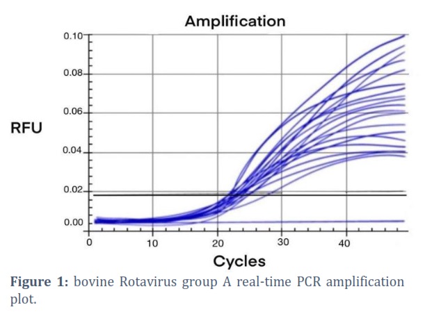
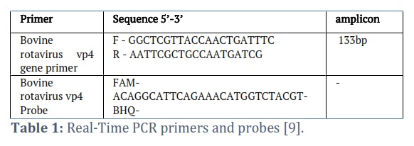
Fecal suspension was prepared in DEPC water to 1:4 dilution then clarified at 5000 rpm\15 minutes at 4°C and the supernatant submitted to RNA extraction. Viral RNA was extracted from stool samples by using AccuZolTM Total RNA extraction kit (Bioneer, Korea) and done according to company instructions. The extracted RNA sample was kept at -20 freezers. The extracted RNA from stool samples were estimated by using Nanodrop spectrophotometer that used to measurement the RNA concentration and purity at absorbance 260/280 nm at ratio 1.8 as pure RNA, The RNA samples were used in cDNA synthesis step by using M-MLV Reverse Transcriptase kit and done according to company instructions. RNA and primer were denatured for 10 min at 65 °C, after that immediately cool on ice Then the tubes were placed in vortex and briefly spinning down. The RNA converted into cDNA in thermocycler qPCR was performed for detection of bovine rotavirus based on VP4 gene.
Blood samples
Jugular vein blood samples were taken from buffalo calves that tested positive for rotavirus. A blood sample of ten milliliters was taken. A test was conducted to determine whether these calves have an infection. The blood samples were collected using syringes that had a volume of 10 milliliters. Following the processing of the blood samples, each syringe was halved. After the clotting process was finished, the samples were centrifuged after twenty minutes of room temperature. When the blood had clotted, this was finished. After that was finished, the samples were centrifuged again for additional separation. A blood picture was obtained by inserting 3 milliliters of blood into an anticoagulant tube (EDTA), and serum for the chemical tests was collected by inserting 5 milliliters of blood into a plain tube. Blood samples were collected in each of these procedures. The necessary samples were collected by means of each of these tubes. A control group of six healthy calves had their blood samples obtained in the same manner as the infected group so that the two groups could be compared. Each of the two data sets was compared. The purpose of this was to highlight the dissimilarities between the two groups. Performing this task was supposed to accomplish the objective of providing a comparison between the two sets. Using serum as a measuring instrument, a dry chemical analyzer can determine the concentrations of salt and potassium in a sample.
Results![]()
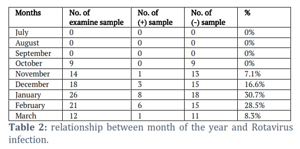
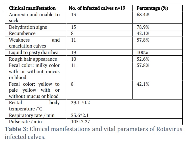
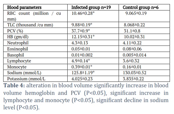
Out of 100 fecal samples from buffalo calves suffered from diarrhea, Rotavirus infection detected in nineteen (19) samples via rotavirus antigen rapid test kit as a field diagnosis. Nineteen positive samples confirmed by real time PCR, yielding infection rate was 19%. there’s no significant difference between male and female calves rotavirus infection. Rotavirus infection at the age group(1-4) day old was 4 from 27 samples (14.8%), while at age group (5-14) the highest rate of Rotavirus was (27.5%) 8 positives from all 29 samples, age group(15-21) gave 5 positive samples from 23 (21.7%) and the lowest infection rate was recorded at age group(21-30) which was (9.5%) 2 positive sample of all 21 Diarrheic buffalo calves. With reference to infection rotavirus according to the months of the year table (2) was 1 positive rotavirus from 14 Diarrheic fecal samples (7.1%) at November, from 26 samples there were 6 positives(16.6%) at the December, At January month recorded highest rotavirus infection rate(30.7%) 8 positives from 26 samples, (28.5%) infection rate was in February month 6 positives from 21 samples and at March month recorded 1 positive from 12 samples (8.3%). The months from July to September there wasn’t recoded any case of diarrhea in buffalo calves, on October 9 fecal samples were examined gave negative results for rotavirus infection. Clinical examinations on rotavirus infected calves revealed depression anorexia and dehydration (loses of skin elasticity and sunken eyes)skin fold test in dehydrated calves showing Mild to Moderate and Severe dehydration signs. In addition, there is variation in rectal body temp, pulse rate and respiration rate(39.1±0.2), (38.8±0.1) rectal body temperature of infected and control group respectively. Respiratory rate was(23.6±2), (25.3±0.93) in infected and control group respectively and pulse rate was (105±2.27), (107.5±0.69) in infected and control groups respectively as shown in table (3). There’s elevation(P<0.05) in RBC volume, PCV and hemoglobin also significantly increase(P<0.05) in TLC(10.46±0.28), (37.7±0.9),(12.15 ±03) and (9.88 ±0.19) respectively. there was significant increase (P<0.05) in lymphocytes and monocytes (4.9 ±0.14), (0.39±0.01) respectively. A significant difference has been encountered in biochemical analysis of infected buffalo calves than control group, significance(P<0.05) decrease was indicated in infected buffalo calves than control group, there were no significant (P<0.05) deference between infected and control group in potassium as shown in table (4).
Figures & Tables
Increased viral shedding from stools and irritable bowel syndrome are two signs of rotavirus infection. Rotavirus infection manifests itself in a variety of ways. There is a chance that it can lead to financial losses since these symptoms are linked to rotavirus infection. The small intestine's cells, known as enterocytes, are susceptible to viral infection, which may lead to diarrhea [10]. It is this virus that causes the sickness. Diarrhea is the end result of this virus's assault on enterocytes. There have been recent episodes of diarrhea, and this virus is to blame.
The rapid and accurate diagnosis of bovine rotavirus (BRV) infection in buffalo calves in Basrah Governorate is crucial for disease control and eradication, particularly in newborn animals on buffalo farms. Rapid diagnosis of BRV is essential for initiating appropriate therapy and avoiding unnecessary antibiotic use. Traditional diagnostic methods involving RNA extraction and conversion to cDNA are labor-intensive and time-consuming. However, rapid test kits can swiftly and accurately detect prevalent enteropathogens associated with diarrhea in newborn calves [11]. In this study, all 19 positive samples tested using a rapid test kit confirmed rotavirus infection. Our findings regarding rotavirus infection in buffalo calves (19%) closely align with a previous study (18.18%) [12] but differ significantly from another study (4.76%) [13]. Males showed a higher infection rate (21.17%) compared to females (16.6%), although the difference was not statistically significant. This study aligns with a previous report [14] but contradicts another [10]. The risk of BRV-A infection in calves peaks during the first month of life and decreases with age, likely due to neutralization of digestive tract acidity by suckling milk, enabling pathogen survival [15]. Our investigation revealed the highest infection rates in calves aged 5-14 days (27.5%) and 15-20 days (21.7%), with the lowest rate in the 21-30-day group(9.5%), consistent with prior research [16]. BRV-A infections were highest in January (30.7%), followed by February (28.5%), December (16.6%), and March (8.3%), with the lowest rate in November (7.1%). Summer and early autumn showed no recorded cases, likely due to the calving season occurring from late autumn to spring [17,18]. This trend is consistent with other studies indicating a winter peak in rotavirus-caused diarrhea, correlating with adverse weather conditions [19].
Clinically, BRV infection in buffalo calves causes diarrhea leading to dehydration, metabolic acidosis, muscle weakness, anorexia, and hypothermia [20]. Dehydration and metabolic acidosis contribute to increased rectal temperatures due to reduced heat elimination efficiency, followed by a decrease in body temperature. Metabolic acidosis stimulates the sympathetic nervous system, leading to elevated heart rates (tachycardia) and respiratory rates (hyperpnea) [21,22]. Elevated RBC count, hemoglobin, PCV, and WBC count, indicating hemoconcentration due to dehydration and metabolic acidosis [23]. Elevated white blood cells suggest an active immune response against infection, particularly monocytes involved in innate immunity and cytokine production[26,27]. Diarrhea in BRV-A-infected calves causes electrolyte imbalances and dehydration due to increased fecal electrolyte loss and intestinal nutrient malabsorption [28,29]. However, our study did not observe hyperkalemia but noted a significant decline in serum sodium levels due to direct fecal sodium loss and reduced intestinal absorption, consistent with previous findings [23] but contradictory to others [30].
Acknowledgement
Current study was performed under the permission of the ethical committee in the faculty of veterinary medicine university of Basrah (Ref. No. 77/2022).
Conflict of Interest
The authors declare that there is no conflict of interest regarding the publication of this paper.
Asma Salim: molecular detection, field diagnosis of the disease and manuscript writing.
Muhsen R.K: supervision, mythology study design, data curation.
![]() References
References
- Bertoni E, Barragán AA, Bok M, Vega C, Martínez M, et al. Assessment of influential factors for scours associated with cryptosporidium sp., rotavirus and coronavirus in calves from argentinean dairy farms. Animals, (2021);11(9): 2652.
- Lee SH, Kim HY, Choi EW, et al. Causative agents and epidemiology of diarrhea in Korean native calves. Journal of Veterinary Science, (2019);20(6).
- Miño S, Kern A, Barrandeguy M, et al. Comparison of two commercial kits and an in-house ELISA for the detection of equine rotavirus in foal feces. Journal of Virological Methods, (2015); 222: (1-10).
- Sawant P M, Digraskar S S, Gopalkrishna V. Molecular characterization of unusual G10P [33], G6P [14] genomic constellations of group A rotavirus and evidence of zooanthroponosis in bovines. Infection, Genetics and Evolution, (2020); 84: 104385.
- Heredia N, García S. Animals as sources of food-bornepathogens: A review. Animal Nutrition, (2018); 4(3): 250-255.
- Soltan MA, Tsai YL, Lee PYA, et al. Comparison of electron microscopy, ELISA, real time RT-PCR and insulated isothermal RT-PCR for the detection of Rotavirus group A (RVA) in feces of different animal species. Journal of virological methods, (2016); 235: (99-104).
- Kassem IK, Magouz AF, Desouky AY, et al. Isolation and Identification of Rotavirus Infection in diarrheic calves at El Gharbia Governorate. Global Veterinaria, (2017); 18 (3): 178-182.
- Aldawmy FK, Thwiny HT, Abo Almaali HM. Epidemiological and molecular study of Rotavirus infection among human and animal in Karbala and Basrah provinces. Iraqi Journal of Veterinary Sciences, (2021); 35(2): 403-410.
- Hardy ME, Woode GN, Xu ZC, et al. Comparative amino acid sequence analysis of VP4 for VP7 serotype 6 bovine rotavirus strains NCDV, B641, and UK. Journal of virology, (1991); 65(10): 5535-5538.
- Makwana PM, Kalyani IH, Desai D,et al. Detection of bovine rotavirus (BRV) infection in neonatal calves of in and around Navsari district of South Gujarat, India. J Entomol Zool Stud. 2020a, (2020); 8(2): 1092-7.
- SAKLI GU, Bulut O, Hasöksüz M, et al. Investigation of bovine coronavirus and bovine rotavirus by rapid diagnosis kit and RT-PCR in diarrheic calf feces. Journal of Istanbul Veterinary Sciences, (2019); 3(3): 57-63.
- Sharma SK, Monika J. Epidemiological studies on calf diarrhoea. Indian Veterinary Journal, (2018); 95(3): 32-4.
- Udaykar A, Sharda R, Malik Y S, et al. Outbreak of calf diarrhea due to rotavirus infection and an assessment of the economic impact. Tropical Animal Health and Production, (2019); 51: 1861-1867.
- Patel J, Mathakiya R, Golaviya A. Detection of bovine rotavirus from diarrheic bovine calves in Gujarat region, India. International Journal of Current Microbiology and Applied Science, (2019); 8(9): 1282-1293.
- Lora I, Gottardo F, Contiero B, et al. Association between passive immunity and health status of dairy calves under 30 days of age. Preventive veterinary medicine, (2018); 1(152): 12-5.
- KS KA. Detection of Rota and Corona viral antigens in diarrheic newly born calves in Menofiya governorate. Benha Veterinary Medical Journal, (2015); 29(1): 9-16.
- Al-Delemi DH, Kadhim MS. Effect of season on the some blood hormones and enzymes of Iraqi bull buffalo.Journal of Dairy, Veterinary & Animal Research, (2015); 2(5):10-53.
- Al-Yasiri EA. Estimation of Progesterone and Estradiol in Blood Serum in Iraqi Buffaloes Related with Normal Parturition and Dystocia. Biochemical & Cellular Archives, (2021); 1(21): 1.
- Berber E, Çanakoğlu N, Sözdutmaz İ, et al. Seasonal and age-associated pathogen distribution in newborn calves with diarrhea admitted to ICU. Veterinary Sciences, (2021);98(7): 128.
- Radostits O M, Gay C C, Hinchcliff K w, et al. Chapter one: Veterinary. Medicine. A text book of disease of Cattle, sheep, pigs, & horse. (2007); 10th ed: 75. Saunders Elsevier, Edinburgh.
- Constable PD, Hinchcliff KW, Done SH, et al. Veterinary medicine: a textbook of the diseases of cattle, horses, sheep, pigs and goats. Elsevier Health Sciences(2016);1(11):67-77.
- El-sheikh AK, Hayam M, Morsy S, et al. Clinical and laboratory examinations of diarrhea and dehydration in newborn Friesian calves with special reference to therapy with hypertonic and isotonic solution. Life Science Journal, (2012);9(4): 181-4.
- mohammed Abed ZE, Sulaiman YA, Khalaf HY. Evaluation of some biochemical and Hematological parameters for Changes Associated with Diarrhea in Calves. Tikrit Journal of Pure Science, (2020); 28;25(1): 7-9.
- Shekhar S, Ranjan R, Singh CV, et al. Prevalence, clinicohaemato-biochemical alterations in colibacillosis in neonatal calves. International Journal of Current Microbiology and Applied Science, (2017);6(9): 3192-3198.
- Kim HJ, Park JG, Matthijnssens J, et al. Intestinal and extra-intestinal pathogenicity of a bovine reassortant rotavirus in calves and piglets. Veterinary microbiology, (2011); 152(3-4): 291-303.
- Kim S, Yu DH, Jung S, et al. Biological Factors Associated with Infectious Diarrhea in Calves. Pakistan Veterinary Journal, (2021); 1(41): 4.
- Abdulkhaleq LA, Assi MA, Abdullah R, et al. The crucial roles of inflammatory mediators in inflammation: A review. Veterinary world, (2018);11(5): 627.
- Sayers RG, Kennedy A, Krump L, et al. An observational study using blood gas analysis to assess neonatal calf diarrhea and subsequent recovery with a European Commission-compliant oral electrolyte solution. Journal of Dairy Science, (2016);1;99(6): 4647-55.
- Seifi HA, Mohri M, Shoorei E, et al. Using haematological and serum biochemical findings as prognostic indicators in calf diarrhoea. Comparative Clinical Pathology, (2006); 15: 143-7
- Alkhafaji I J and Mahmood AK. “ The study of biochemical ions parameters of non-diarrheic and diarrheic Iraqi Awassi suckling lambs.” Al-Anbar journal of veterinary Sciences (2016);9(2): 110-113.
This work is licensed under a Creative Commons Attribution-Non Commercial 4.0 International License. To read the copy of this license please visit: https://creativecommons.org/licenses/by-nc/4.0


