

Full Length Research Article
Segmental Structure of the Human Kidney: An Innovative Research Algorithm
Edgar Kafarov*, Imran Avduev, Zelimkhan Lechiev
Adv. life sci., vol. 10, no. 4, pp. 549-554, December 2023
*- Corresponding Author: Edgar Kafarov (kafarov.e.s@mail.ru)
Authors' Affiliations
[Date Received: 24/07/2023; Date Revised: 05/09/2023; Date Published: 31/12/2023]
Abstract![]()
Introduction
Methods
Results
Discussion
References
Abstract
Background: Understanding the anatomical structure of the human kidney, particularly its zonal and segmental organization, is crucial for both theoretical knowledge and practical applications in surgical procedures. That is why the purpose of the article was to study the variants of the zonal and segmental structure of human kidneys.
Methods: 116 corrosive preparations of the arterial system of the human kidney served as the material for the study. The authors identified their extra-organ branches: a) the number of arterial vessels in the kidney hilum; b) the topographic and anatomical features of the renal artery trunks. In a 3D projection, zones of local zonal blood supply to areas of the renal parenchyma were identified, depending on the di- and trichotomic variants of the division of the main renal artery, A. renalis (I).
Results: It was found that some lines of the passage of the zones of natural divisibility of the kidneys with different variants of the division of A. renalis (I). There were from 5 to 7 (6 ± 1) segments in the kidneys on average. It is possible that if a four-zone blood supply system with corresponding zones of natural kidney divisibility is found in the kidneys, where the zones mostly don’t coincide with the boundaries of the passage of segments in their classical version, the number of segments will be even greater.
Conclusion: The results of the conducted study show that considering the generally accepted zones and boundaries of the passage of segments with zones of natural divisibility, which were not previously considered, the kidneys have an individual segmental structure, which requires revision. The authors believe that the generally accepted classical five-segment kidney model is outdated.
Keywords: Kidney; Artery; Vein; 3D Analysis
Introduction![]()
Many researchers have written on the issues of angio-architectonics and the segmental structure of the kidney [1-4].There is still no unified point of view about the lobular, zonal, and segmental structure of the kidney, and the main criteria used for dividing this organ into fractions or segments have not been completely defined [5-7]. Knowledge of the variants of the segmental structure of the kidneys is of great importance in practical surgery for performing organ-preserving operations or segmental resections [8-10]. By the division of the branches of the main renal artery, A. renalis (I),the kidney is divided into segments which are called arterial segments of the kidney [11]. In modern science, the classical five-segmental structure of the kidney is generally accepted, where the superior, superior anterior, inferior anterior, inferior, and posterior segments, i.e., isolated areas of the kidney parenchyma supplied with blood by pools of segmental arteries, are distinguished. As a result, five segmental arteries define the segments of the kidney, as in principle, it is depicted in many literary publications on human anatomy, including atlases on human anatomy [12]. There are many works in the literature devoted to the segmental structure of the kidneys, since this is closely related to the question of the presence of low-vascular zones in the kidneys and to determine the segmentation of the organ, the arterial vessel is isolated as the main morphological feature [13,14]. However, according to the studies of these authors, the number of segments in the kidneys is not the same [15, 16]. This is probably because the authors take branches of different orders for the segmental artery [17,18]. The International Anatomical Nomenclature (2003) in the arterial bed of the kidney does not contain such a term as a segmental artery. A branch of the 2nd or 3rd order is usually called a segmental artery. If it is a branch of the 2nd order, then this is the result of the division of A. renalis (I), that is, the first level of division. According to some authors, in most cases, A. renalis (I) is divided into two branches, ventral and dorsal [19,20]. It should be noted that the kidney segments were studied by the authors mainly with the variants of the division of A. renalis (I) into ventral and dorsal branches, not considering other possible variants of the division of the renal artery and the types of intra-organ branching.
There is also the issue of the sources of formation and the number of segmental arteries in different kidneys. If these are branches of the 3rd order, then this is the third level of division, interlobular arteries located in the parenchyma of the organ, the number of which can reach from 4 to 10, i.e.,10 segmental arteries. However, according to the literature review and International Anatomical Nomenclature, there are only five segments, which require specification [21]. In classical textbooks and atlases of human anatomy, among other things, a five-segment model of the kidney is illustrated, where the division of A. renalis (I) into ventral and dorsal branches is mainly depicted [22,23]. Further, the ventral branch of the renal artery, as illustrated in a textbook or an atlas, is divided into arterial vessels that supply blood to the segments of the kidney. Moreover, the ventral branch, being the maternal one, regardless of the type of branching (magistral or dispersed), splits into four arteries of the 3rd order, that is, segmental arteries going to the four renal segments [24,25]. However, the dorsal branch, as shown in many textbooks and atlases, smoothly passes into the dorsal segmental artery for blood supply to the only posterior segment of the kidney. Although the dorsal branch of the renal artery, as well as the ventral one, also splits into several branches of the 3rd order, based on the principles of di- or trichotomy, they should be called interlobular or segmental arteries. In the dorsal artery, as in the ventral one, each 3rd-order interlobular artery or segmental artery in the renal parenchyma also forms its arterial vascular pool, which has a certain isolated area of blood supply to the renal parenchyma [26]. However, this pattern does not apply to the dorsal artery, which requires a separate specification.
This gap is not the only one in the literature among many groups of questions that have arisen concerning the specifics of the structure of the human kidney bloodstream. Even the information about the main renal vessels seems to be contradictory in some respects [27]. In our opinion, this is due to the countless individual variability of the topography of the renal vessels and the shape of the intra-organ branching of blood vessels, both within the hilum and sinuses of the kidneys, and in the thickness of their parenchyma. Based on this, the relevance of this problem becomes obvious. The purpose of this study is to review the variants of the zonal and segmental structure of human kidneys.
Methods![]()
Research design
The study used a descriptive design to investigate the segmental structure of the human kidney and determine the zones of natural divisibility. We used a combination of experimental and imaging techniques to analyze arterial preparations and 3D models of the renal arterial bed.
Research materials
We used 116 corrosive preparations of the arterial system of the human kidney made of fast-hardening polymers.
Research algorithm
- The finished preparations were scanned [20] and photographed using a Sony Cyber-shot DSC-RX10M 4 Black digital camera.
- On corrosive preparations of arterial vessels of the kidneys and 3D scans of renal arteries in 3D projection, their extra-organ branches were observed:
a) the number of arterial vessels in the kidney hilum;
b) topographic and anatomical features of the trunks of the renal arteries in the kidney hilum.
- In the Mimics 8.1 software, 3D models of the renal arterial bed showed a 3D projection of the main arteries of the kidney relative to different planes (frontal, horizontal, and sagittal).
- In a 3D projection, zones of local blood supply to areas of the renal parenchyma were identified, depending on the di- and trichotomic variants of the division of A. renalis (I). Based on the analysis of literary sources, it was accepted that certain places in the parenchyma of the kidneys, where small branches of arterial vessels of the basins of zonal arteries, A. zonal (II),met but did not intersect or did not cross their boundaries, were called zones of natural divisibility of the kidneys.
- In a computer program, by combining 3D images of a classical five-segment kidney model with a 3D image of the obtained variants of natural divisibility zones, their multimodal images were created [28]. A geometric correction was carried out by projective transformations.
Data analysis
All obtained digital materials and data from instrumental research methods were processed based on variation statistics methods using a workstation with an Intel Core2Duo T5250 1.5 Ghz processor and 2GB RAM on the Windows 7 platform. In the course of the work, we used the Excel application package from Microsoft Office 2007.
Results![]()
As a result of creating multimodal images of combined images of the classical five-segment kidney model with six variants of the topography of the zones of their natural divisibility, new variants of the segmental structure of the human kidney were obtained.
When presenting the results of our study, we used the generally accepted classical definitions of some anatomical formations of the kidneys based on the analysis of literary sources for convenience. Thus, under the kidney zone, we understood a section of the renal parenchyma, in which a branch of the renal artery of the 2nd order was distributed, that is, A. zonal (II). The topography of A. zonal (II) in the kidney hilum, their features of intra-organ branching, and the formation of arterial basins in various areas of the renal parenchyma depend on the options for passing zones of natural divisibility on the surface of the kidney.
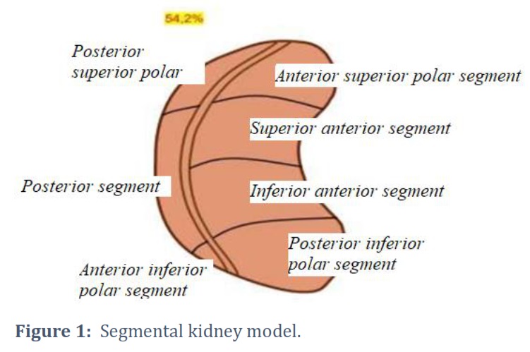
By the segment of the kidney, we understand a section of the parenchyma where the branch of the 3rd order divides, that is, the vessels of the interlobar artery basin, A. interlobares – 1 (III) or the segmental artery [29]. When creating multimodal images, it was found that some segments of the kidney were still divided into parts by zones of natural divisibility, depending on the variants of the division of A. renalis (I) into A. zonal (II). Thus, in the 1st variant of the division of A. renalis (I) into ventral and dorsal A. zonal (II), which was observed in 54.2% of cases. The zone of natural divisibility of the kidney ran along its lateral edge, then starting from the upper corner of the renal sinus it was directed upwards, then it was directed along the superior pole of the kidney, dividing the superior polar segment into two more segments. Then it descended to the inferior pole of the kidney, dividing this segment into two more segments, retreating 0.5 cm posteriorly from its convex edge. In this variant, the kidney had the following segments (Figure 1).
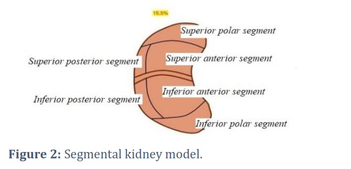
In the 2nd variant of the division of A. renalis (I) into superior polar and inferior polar A. zonal (II), which was observed in 15.5% of cases, the zone of natural divisibility of the kidney ran along the anterior surface of the kidney from its medial edge and the middle of the anterior lip to the lateral edge of the kidney. Then it passed through the convex edge of the kidney to the posterior surface, dividing the posterior segment into two more segments and reaching the medial edge. In this variant, the kidney had the following segments (Figure 2).
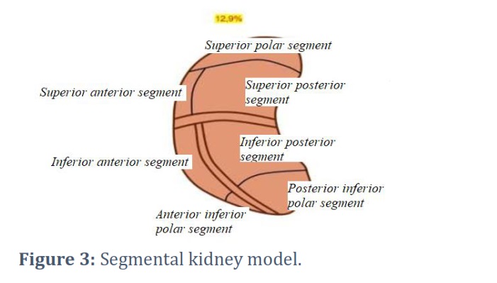
In the 3rd variant of the division of A. renalis (I) into ventral, dorsal, and superior polar A. zonal (II), the first zone of natural divisibility of the kidney passed along the anterior surface of the kidney in a transverse direction, from its medial edge and the middle of the anterior lip to the lateral edge of the kidney. Then it passed through the convex edge of the kidney to the posterior surface, dividing the posterior segment into two more segments and reaching the medial edge. The second zone of natural divisibility of the kidney began from the first one, retreating 0.5 cm posteriorly from the lateral edge of the kidney, heading down along its convex edge, dividing the inferior polar segment into two more segments and reaching the lower corner of the kidney hilum. In this variant, the kidney had the following segments (Figure 3).
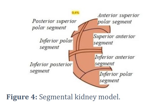
In the 4th variant of the division of A. renalis (I) into ventral, dorsal, and inferior polar A. zonal (II), which we found in 9.4% of cases, the first zone of natural divisibility of the kidney in this variant passed along the anterior surface of the kidney in the transverse direction, from its medial edge and the middle of the anterior lip to the lateral edge of the kidney. Then it passed through the convex edge of the kidney to the posterior surface, dividing the posterior segment into two more parts and reaching the medial edge. The second zone of natural divisibility of the kidney began from the first one, retreating 0.5 cm posteriorly from the lateral edge of the kidney, went up along its convex edge, reached the superior polar segment, dividing it into two parts, and reached the superior corner of the kidney hilum. In this variant, the kidney had the following segments (Figure 4).
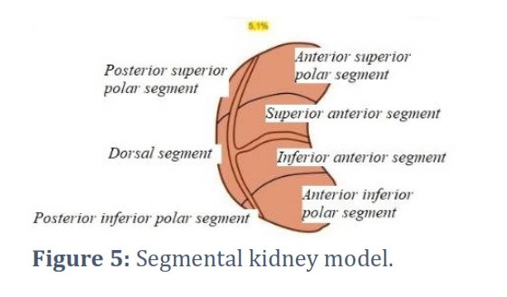
In the 5th variant of the division of A. renalis (I) into two ventral and one dorsal A. zonal (II), which we found in 5.1% of cases. The first zone of natural divisibility of the kidneys in this variant passed longitudinally along its lateral edge, then starting from the upper corner of the renal sinus it went up, then along the superior pole of the kidney, it descended to the inferior pole, dividing the superior polar segment and the inferior polar segment into two more parts, retreating 0.5 cm posteriorly from its convex edge and closing in the lower corner of the renal sinus. The second zone of natural divisibility of the kidney passed in the transverse direction, corresponding to the boundary between the superior anterior and inferior anterior segments. On the dorsal surface, the zone of natural divisibility also started from the central sections of the longitudinal line, then went in a transverse direction towards the kidney hilum. In this variant, the kidney had the following segments shown (Figure 5).
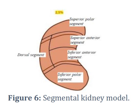
In the 6th variant of the division of A. renalis (I) into superior polar, central, and inferior polar A. zonal (II), which we found in 2.5% of cases, the first zone of natural divisibility of the kidney, passing from the central parts of the kidney hilum, starting from the anterior lip and heading laterally, corresponded to the border between the superior polar and the superior anterior segments. The second zone of natural divisibility of the kidney also passed from the central parts of the kidney hilum, starting from the anterior lip, then going laterally in an oblique downward direction and corresponding to the border between the inferior anterior and inferior polar segments. On the dorsal surface of the kidneys, both zones of natural kidney divisibility in this variant corresponded to the boundaries between the superior polar, dorsal, and inferior polar segments. In this variant, the kidney had the following segments (Figure 6).
Figures & Tables
Thus, studies show that only in the case of the division of A. renalis (I) into the superior polar, central, and inferior polar A. zonal (II), which was observed in 2.5% of cases, the kidney has a classic five-segment structure [30]. However, in our opinion, this percentage is a small indicator to bring on average all the kidneys to the generally accepted five-segment structure. In the case of the most common variant of the division of A. renalis (I) into ventral and dorsal A. zonal (II), which was found in 54.2% of cases, the kidney has seven segments. On average (X ± m) there are from 5 to 7 (6 ± 1) segments in the kidneys, which to some extent corresponds to the data of Domnitsky et al., and Shahul Hameed et al., [31, 32]. It is possible that in the case of a four-zone blood supply system with corresponding zones of natural kidney divisibility, where the zones do not coincide with the boundaries of the passage of segments in their classical version, their number will be even greater [33].
Thus, the results of studies conducted on a sufficient amount of material, considering the generally accepted zones and boundaries of the passage of segments with zones of natural divisibility of the kidneys, which were not previously considered [34], show us that the kidney, depending on the variants of the division of A. renalis (I) into A. zonal (II), with the formation of their arterial basins and considering the zones of natural divisibility has an individual segmental structure, which requires a revision [35]. We cannot agree with the generally accepted classical five-segment kidney model and consider it outdated.
Our study is based on a 3D analysis of the variants of the division of A. renalis (I) where six variants of its division into A. zonal (II), forming arterial vascular pools in the parenchyma of the kidney with the formation on the surface of the kidneys of variants of the passage of zones of their natural divisibility. We found that in 54.2% of cases, A. renalis (I) is divided into ventral and dorsal A. zonal (II), relative to the frontal plane, forming the ventral and dorsal zonal blood supply systems of the kidneys (bi-zonal blood supply system of the kidneys). In 15.5% of cases, A. renalis (I) is divided relative to the horizontal plane into the superior polar and inferior polar A. zonal (II), forming the superior polar and inferior polar zonal blood supply systems in the kidneys (bi-zonal kidney blood supply system).
Further, variants of kidneys with a three-zonal blood supply system were identified: for example, in the 1st variant, A. renalis (I) in 12.9% of cases, was divided relative to the frontal and horizontal plane into superior polar, ventral, and dorsal A. zonal (II). In the 2nd variant, A. renalis (I) was divided relative to the frontal and horizontal plane into ventral, dorsal and inferior polar A. zonal (II), which was observed in 9.4% of cases. In the 3rd variant, which was observed in 5.1% of cases, A. renalis (I) was divided relative to the frontal plane into two ventral and one dorsal A. zonal (II).In the 4th variant, A. renalis (I) was divided into the superior polar, central, and inferior polar A. zonal (II), which was observed in 2.5% of cases. The variants of zonal blood supply to the kidneys that we identified formed zones of natural divisibility on their surface.
Conflict of Interest
The authors declare no conflict of interest.
The research presented in this study is a collaborative effort that reflects the equal contributions of authors. All authors participated actively in conceiving and designing the study, as well as in the acquisition, analysis, and interpretation of the data.
![]() References
References
- Chen G, Dai Y, Li R, Zhao Y, Cui L, Yin X. SDFNet: Automatic segmentation of kidney ultrasound images using multi-scale low-level structural feature. Expert Systems with Applications, (2021); 185: 115619.
- Spiegel M, Hahn DA, Daum V, Wasza J, Hornegger J. Segmentation of kidneys using a new active shape model generation technique based on non-rigid image registration. Computerized Medical Imaging and Graphics, (2009); 33(1): 29–39.
- Rombolotti M, Sangalli F, Cerullo D, Remuzzi A, Lanzarone E. Automatic cyst and kidney segmentation in autosomal dominant polycystic kidney disease: Comparison of U-Net based methods. Computers in Biology and Medicine, (2022); 146: 105431
- Sun P, Mo Z, Hu F, Song X, Mo T, et al. Segmentation of kidney mass using AgDenseU-Net 2.5D model. Computers in Biology and Medicine, (2022); 150: 210-221.
- Breshears MA, Confer AW. The Urinary System. Pathologic Basis of Veterinary Disease, (2017); 617–681.
- Blanc T, Goudin N, Zaidan M, Traore MG, Bienaime F, Turinsky L, Garbay S, Nguyen C, Burtin M, Friedlander G. Three-dimensional architecture of nephrons in the normal and cystic kidney. Kidney International, (2021); 99: 632–645.
- Little MH. Returning to kidney development to deliver synthetic kidneys. Developmental Biology, (2021); 474: 22–36.
- Gianto EK. Characteristics of COVID-19 Patients who Developed Acute Kidney Injury and Its Association with Mortality: A Systematic Review. Gaceta Médica De Caracas, (2023); 131(S2): 137-143.
- Sysoev PA, Vezirkhanov AZ, Kafarov ES. Variant Anatomy of Blood Supply Sources of Human Kidney Segments. Journal of Morphological Sciences (2022); 39:521–528.
- Malzoni M, Iuzzolino D, Rasile M, Coppola M, et al. Surgical Principles of Segmental Rectosigmoid Resection and Reanastomosis for Deep Infiltrating Endometriosis. Journal of Minimally Invasive Gynecology, (2020); 27(2): 258.
- Kafarov ES, Asfandiyarov FR. Branching types of arterial and venous vessels of the kidney. Morphological statements (2008); 3(4):41–42.
- Kafarov ES, Asfandiyarov FR. Clinical and anatomical aspects of the topography of the renal artery, vein and pelvis. Rossiiskie morfologicheskie vedomosti, (2008); 3:3–4.
- Ray N, Reddy PH. Structural and physiological changes of the kidney with age and its impact on chronic conditions and COVID-19. Ageing Research Reviews, (2023); 88: 1-8.
- Yoshida K, Takamatsu A, Nohara T, Yoneda N, Inoue D, et al. Renal artery-based kidney segmentation on CT for patients with renal cell carcinoma: Feasibility of segmental artery clamping simulation. European Journal of Radiology Open (2023); 10: 1-5.
- Ruan Y, Hong F, Lin M, Wang C, Lian F, et al. Clinicopathological characteristics, risk factors and prognostic value of intrarenal vascular lesions in IgA nephropathy. European Journal of Internal Medicine (2023); 117: 91-97.
- Kuan JK, Wright JL, Nathens AB, Rivara FP, Wessells H. American Association for the Surgery of Trauma organ injury scale for kidney injuries predicts nephrectomy, dialysis, and death in patients with blunt injury and nephrectomy for penetrating injuries. Journal of Trauma – Injury, Infection and Critical Care (2006); 60(2): 351-356.
- Kvyatkovskaya TA, Chernyavskii EKh, Kutsyak TL. Anatomical and sonographic comparison of morphometric data of renal vessels and their intraorganic branches. Rossiiskie morfologicheskie vedomosti, (2000); 1–2:201–202.
- Alyaev YuG, Sirota ES, Bezrukov EA, Ali SKh. 3D technologies in planning and navigation of laparoscopic operations in patients with kidney and ureter calculi. Urologiya, (2019); 4:9–15.
- Asfandiyarov FR, Kafarov ES. Variant anatomy of the vascular bed of the kidney. Astrakhanskii meditsinskii zhurnal, (2007); 2:2 3.
- Burgener FA, Kormano M, Pudas T. Bone and Joint Disorders : Conventional Radiologic Differential Diagnosis: a manual: an atlas: more than 1000 radiographs. Moscow: GEOTAR Media (2014).
- Kaplunova OA. Possibilities of X-ray angiography and spiral computed angiotomography in the study of extra- and intraorgan renal arteries. Morfologicheskie vedomosti, (2004); 1–2:47.
- Alyaev YuG, Krapivin AA. Site of kidney resection for cancer, In: Promising directions in the diagnosis and treatment of kidney cancer: materials of the research and practice conference, 18. Moscow (2003).
- Alyaev YuG, Sinitsyn VE, Grigorev NA. The use of MRI for the selection of the type and volume of surgery for a kidney tumor, In: Promising directions in the diagnosis and treatment of kidney cancer: materials of the research and practice conference, 28. Moscow (2003).
- Glybochko PV, Alyaev YuG, Ternovoy SK, Dzeranov NK, Akhvlediani ND, et al. Computer modeling: an innovative technique in the diagnosis and treatment planning for patients with surgical kidney diseases. Uralskii meditsinskii zhurnal, (2012); 9: 84–87.
- Glybochko PV, Alyaev YuG, Fiev DN, Khokhlachev SB, Grigoryan VA, et al. Computer modeling in planning percutaneous operations in patients with staghorn nephrolithiasis. Saratovskii nauchno-meditsinskii zhurnal, (2011); 5: 137.
- Areej H, Farooq SMY, Asad N, Fatima M, Mahmood A, Attique UK, Tahir MU, Khan HN. Carotid artery Disease Assessed by Color Doppler Flow Imaging: Comparison Between Diabetic and Non-Diabetic Patients. Advancements in Life Sciences, (2023); 10:66–71.
- Xu H, Li J, Liu J, Zhang B, Wang W. Clinical experience of minimally invasive duodenum preserving pancreatic head resection. Intelligent Surgery, (2022); 5:12–15.
- Gavrilov N, Belokamenskaya A. Organization of stream computing on GPU in the issue of tomogram stereovisualization, In: High-Performance Parallel Computing on Cluster Systems: Proceedings of the 10th International Conference, 58-61. Perm (2010).
- Amparore D, Piramide F, Pecoraro A, Verri P, Checcucci E, et al. Identification of Recurrent Anatomical Clusters Using Three-dimensional Virtual Models for Complex Renal Tumors with an Imperative Indication for Nephron-sparing Surgery: New Technological Tools for Driving Decision-making. European Urology Open Science, (2022); 38: 60–66.
- Alyaev YuG, Grigoryan ZG, Levko AA. Bilateral asynchronous kidney cancer. Onkourologiya, (2010); 2: 14–21.
- Domnitsky IJ, Demkin GP, Salautin VV, Pudovkin NA. Morphometric characteristics of pathological processes in the lungs, liver and kidneys in calves with catarrhal bronchopneumonia. Annals of Agri Bio Research, (2019); 24(1): 139–147.
- Shahul Hameed AB, Makinyan LG, Shindiev KA, Aliev R, Apresyan VS, Wessam, AZ. Rheumatoid Forefoot Reconstruction Following Minimally Invasive Surgery and Hoffmann-Clayton Procedure with Administration of Plasma Rich in Growth Factors – A 3-Year Follow-Up: A Retrospective Study. Gaceta Médica De Caracas, (2022); 130(3): 491.
- Chen GP, Zhao Y, Dai Y, Zhang JX, Yin XT, Cui L, Qian J. Asymmetric U-shaped network with hybrid attention mechanism for kidney ultrasound images segmentation. Expert Systems with Applications, (2023); 212: 118847.
- Santos R, Bürgi M, Mateos JM, Luciani A, Loffing J. Too bright for 2 dimensions: recent progress in advanced 3-dimensional microscopy of the kidney. Kidney International, (2022); 102: 1238–1246.
- Alyaev YuG, Fiev DN, Petrovskii NV, Khokhlachev SB. The use of intraoperative navigation in organ-preserving surgical interventions for a kidney tumor. Onkourologiya, (2012); 3: 31–36.
This work is licensed under a Creative Commons Attribution-Non Commercial 4.0 International License. To read the copy of this license please visit: https://creativecommons.org/licenses/by-nc/4.0


