

Full Length Research Article
Isolation and identification of a mycotoxin produced by Aphanoascus terreus
Abeer Mohammed Ali Al-garawyi1, Amal Jameel Kadhim2, Majid Mohammed Mahmood3*
Adv. life sci., vol. 10, no. 4, pp. 537-542, December 2023
*- Corresponding Author: Majid Mohammed Mahmood (majidmahmood93@yahoo.com)
Authors' Affiliations
2. Nasiriya Technical Institution, Southern Technical University – Iraq
3. Department of Biology, College of Science, Mustansiriyah University – Iraq
[Date Received: 30/07/2023; Date Revised: 31/08/2023; Date Published: 31/12/2023]
Abstract![]()
Introduction
Methods
Results
Discussion
References
Abstract
Background: Keratinophilic fungi prefer keratin-rich materials such as; horn, hoof, and beak of birds for the purposes of growth and reproduction, they utilize keratin as a source of carbon. Mycotoxins, which are produced as byproducts by fungi, are dangerous to both human and animal health. This research aims to isolate and identify Aphanoascus terreus fungi from the soil as well as determine their potential to create mycotoxins.
Methods: In January–April 2022, 45 soil samples were randomly collected from southern Iraq to isolation and identification of keratinophilic fungi, the hair bait method and molecular techniques were used also, detection of mycotoxin achieved by TLC technique then experimental injection in vivo.
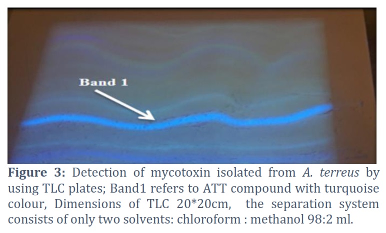
Results: The findings showed that 12 (66.7%) out of 18 soil samples that tested positive for the genus Aphanoascus belonged to the species A. terreus. When these isolates were compared to NCBI using PCR sequencing, they were 99% matched. In addition, all these isolates show the capacity to create a range of unidentified secondary metabolites with a variety of colors and flow rates. Only one compound was studied that appeared with a turquoise hue, so we named it A. terreus T (ATT), which has an Rf. of 18.7 cm. The analysis of secondary metabolites with the aid of FTIR and GC-MS chemical tests indicated possibilities; the most probable is that the ATT is an acidic compound. Visual examinations of the skin of rats injected with ATT showed no obvious abnormalities. Microscopically, they appeared normal as well, but with mild inflammatory signs around the hair follicles.
Conclusion: The outcomes represent the first international registration ever made in accordance with what is known about the production of a mycotoxin from A. terreus. This finding is considered the first reference regarding mycotoxin production from A. terreus.
Keywords: A. terreus; FTIR; Mycotoxins; ATT compound
Introduction![]()
Fungi are a group of organisms that can be found in soil in many places. These living creatures are critical to the soil ecosystem and a source of fungal disease transmission [1]. The skin, hair, nails, fur, feathers, horn, hoof, and beak of birds are examples of keratin-rich materials that keratinophilic fungi prefer for the purposes of growth and reproduction. They utilize keratin as a source of carbon [2]. The phylum Hyphomycetes contains dermatophytes as well as numerous other filamentous fungi. Alternaria, Aspergillus [3], Chrysosporium [4], Cladosporium, Curvularia, Fusarium, Myrothecium, Paecilomyces, Penicillium, Scopulariopsis, and Sepedonium [5] are the most prevalent fungal genera with keratolytic properties. The nuclear ribosomal internal transcribed spacer (ITS) region of DNA has been sequenced the most in fungal molecular ecology research and has been proposed as a universal DNA barcode marker for fungi [6]. Mycotoxins, which are produced as byproducts by fungi, are dangerous to both human and animal health. Mycotoxins have been linked to many problems in the gut, urinary, immune, and reproduction systems [7], and oxidative stress caused by lipid peroxidation has been suggested as a possible cause of mycotoxin-induced harm in these systems [8]. The aim of this research was to isolate and identify A. terreus from soil samples and determine whether it is capable of producing mycotoxins.
Methods![]()
Collection of specimens: In January–April 2022, 45 soil samples were randomly collected from southern Iraq. Sterile disposable spoons were used to gather samples from the surface layer at depths of less than 10 centimeters, and the samples were then put in sterile plastic bags that were labeled.
Criteria for inclusion: agricultural shade areas rich in waste.
Criteria for exclusion: areas directly exposed to sunlight.
In order to isolate keratinophilic fungi, the hair bait method was used [9]. In this case, tufts of horsehair, 1 to 2 centimeters long, were placed over the wet soil samples, and the plates were kept at 270C for 3–4 weeks. The plates were checked often to see if any fungi had grown on the bait. From time to time, distilled water was added to keep the soil moist. Any fungi that grew on the hairs were grown on Sabouraud dextrose agar (SDA) with chloramphenicol, which was used to separate, filter, and identify the fungi.
Conventional identification: According to microbiological guides and atlases, fungal growth was identified based on macroscopically and microscopically observable features [10, 11].
Molecular identification: Adhering to the manufacturer's instructions, genomic DNA was extracted from pure mycelial cultures of the fungal isolates cultivated on SDA using the ZRFungal/Bacterial/Yeast DNA MiniPrepTM Kit (ZYMO, USA). Electrophoresis and Nano-Drop were used, respectively, to evaluate the quantity and properties of the isolated DNA.
Detection of the ITS gene by PCR: The ITS gene was discovered by utilizing primers for amplification. A segment of ITS was amplified using forward and reverse primers (ITS1 F: 5′-TCCG TAGG TGA ACC TG CGG-3′ and ITS4 R: 5′-TCCTCCGCTTATTGATAGATAC-3′) (Integrated DNA Technologies, Canada). PCR amplification was done in a volume of 25 µl contained 1.5µl DNA and 5µl Taq PCR PreMix (Intron, Korea). The Maxime PCR PreMix kit involved the following constituents: 5U/µl i-Taq DNA Polymerase, 2.5 mM DNTPs, 1X reaction buffer (10 X), 1X gel loading buffer, 1µl of each primer (10 pmol) and distilled water were added to complete the volume up to 25µl. Using a thermal cycler (Gene Amp, PCR System 9700; Applied Biosciences), 3 minutes of denaturation at 94°C were followed by 35 cycles of 94°C for 45 seconds, 52°C for 1 minute, and 72°C for 1 minute, with a final incubation at 72°C for 7 minutes. After electrophoresis on 1.5% agarose gels (Conda, USA), the PCR products were seen under UV light (302 nm) following red staining (Intron, Korea). For gene sequencing (Macrogen, Korea), we shipped the PCR products to Korea. Homology was determined using BioEdit and the Basic Local Alignment Search Tool (BLAST), both of which may be found on the NCBI website (http://www.ncbi.nlm.nih.gov).
Detection of mycotoxin production: To test the ability of A. terreus isolates for mycotoxin production, 250 ml Erlenmeyer flasks with Sabouraud Dextrose Broth (SDB) were inoculated and incubated at 28°C for 18 days. Mycotoxin products were extracted according to [12, 13].
Detection of mycotoxin by TLC technique: Thin Layer Chromatography plates (TLC) 20 * 20 cm were used for this approach, which was carried out at the Mycotoxins Laboratory at the College of Applied Medical Sciences/University of Karbala. The TLC plates were activated in an electric oven at 105°C for an hour before use [1415]. Then it was observed under UV light at 365 nm.
Experimental injection in vivo: For the trial, the 12 rats were split up into two groups of six each. The mycotoxin extracted from A. terreus isolates was injected subcutaneously in experimental rats, as has been done by Tovar et al., [16]. Group A: 18 µg\kg of extracted mycotoxin was injected into each rat; group B: 18 µg\kg of normal saline was injected into each rat as a control.
Histopathological examination: Three weeks after performing the injection, the mice were sacrificed, and 1 cm3 of skin samples were taken from the injection area with obvious symptoms. Staining and processing techniques were accomplished according to Suvarna et al., [17].
Fourier Transform Infrared spectrophotometer (FTIR): The detection of functional groups in extracted mycotoxin was done according to Ameer et al., [18] by using the FTIR (Shimadzu) test and ethidium bromide in the chemistry department of the College of Science at Baghdad University.
GC-MS analysis: The identification of chemical compounds in the extracted mycotoxin was done by using a gas chromatography instrument connected to a mass spectrometer (QP210 Ultra, Shimadzu Japan) in the GC-MS laboratory at the Science College, Tehran University, Iran [19].
Results![]()
Morphological identification: As the recent investigation has shown 23 (51.1%) out of 45 soil samples were positive for fungal growth. The most prevalent genus was Aphanoascus, which was isolated from approximately 18 (78.3%) of the positive soil samples, of which 12 (66.7%) belonged to A. terreus.
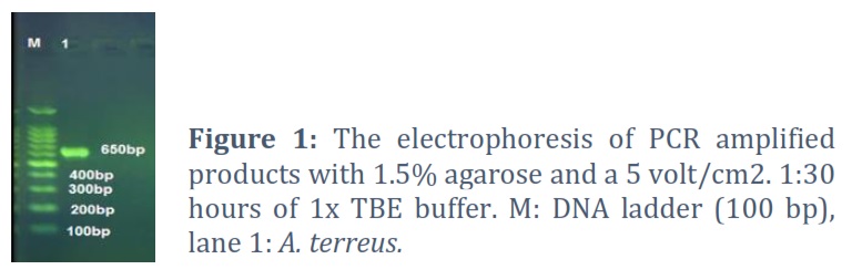
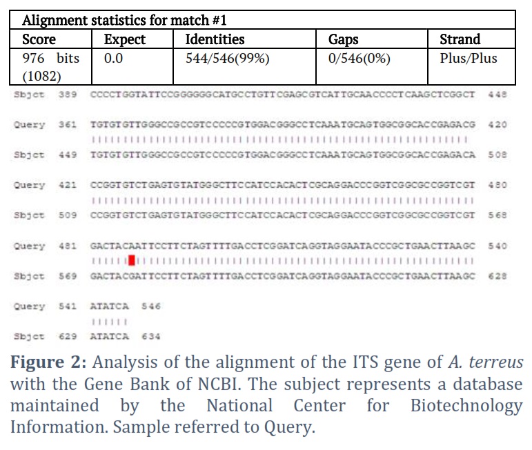
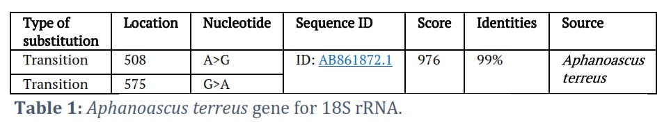
Molecular identification of A. terreus: Different particular genes were used for molecular identification of all fungal isolates in the current study, including 18S rRNA, ITS1, 5.8S rRNA, and ITS2 of 28S rRNA for the 12 A. terreus isolates, by using the universal primers ITS1 and ITS4. Originated a fragment is of approximately 650 bp. The percentage of matching current isolates with NCBI was 99% (Table 1 and Figs. 1 and 2).
Toxicity assay (In vivo): According to the current findings, no laboratory rats died after being subcutaneously injected with mycotoxin ATT compound. As well as, the gross skin examinations of rats showed a normal-like appearance of skin in a compared with control. The microscopic examinations of skin tissue sections after injection with mycotoxin showed mild inflammatory cell infiltrations around hair follicles as compared with the control groups.
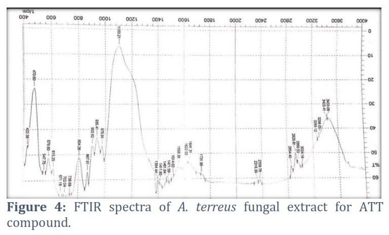
FTIR and GC-MS analysis: The FTIR spectra of A. terreus fungal extract for ATT compound revealed that the band at 3269.12 cm-1 attributed to OH stretching had returned to the carboxylic group COOH. The band around 1471 cm-1 corresponds to the C=C alkene group, whereas 1641.31 cm-1 corresponds to R(C=O) R for carboxylic acid. Furthermore, the band around 1103.21 cm-1 attributed to C-O was returned to carboxylic acid. The result of this test revealed that the ATT compound is likely to be acidic in nature (Fig. 4).
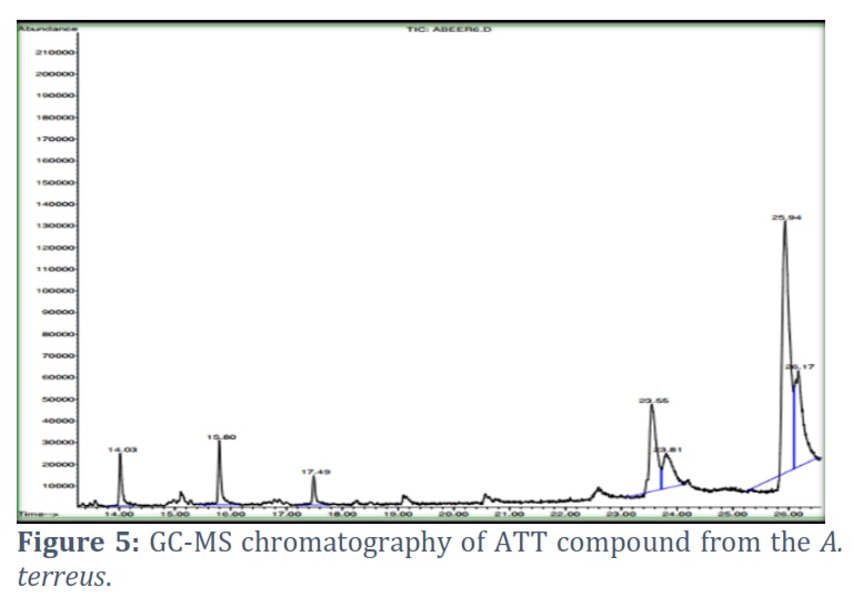
However, the GC-MS chromatography analysis of the A. terreus fungal extract for the ATT compound revealed seven peaks with various possibilities of chemical compounds, most of them acidic compounds Fig.5.
The peak number six had the most likely composition, as follows: 9, 12-octadecadienoic acid (Z, Z) (RT = 25.94 min) having area 42.14 followed by peak number seven showed the possibility: 9,12-octadecadienoic acid (Z, Z)- and (Z)-9-octadecen-4-olide (RT = 26.17 min) with a surface area of 21.52 .
Figures & Tables
As Aphanoascus was found to be the most often occurring genus among the positive soil samples, this is consistent with El-Said [20], who found that Aphanoascus was the most prevalent genus in 26 of 50 isolates (52%). Despite this, Altayyar [21] disclosed that Aspergillus species represented the most abundant (58.9 %) of all isolated keratinophilic fungi, indicating that several species of keratinophilic fungi may exist depending on soil and environment in each country [22]. In addition, the fungal colonies were classified into species by using molecular techniques. The use of these methods allowed for more accurate identification of A. terreus isolates in southern Iraq. This keratinophilic fungus with ovate ascospores and a thick, warty wall is widespread in the world’s soils [23].
In this research all the 12 (100%) A. terreus isolates have the ability to produce various unknown secondary metabolites, no prior research has examined the chemical composition and toxicity of the secondary metabolites isolated from A. terreus, and many different chromatographic methods could be used to quantify them, according to Kizis and his colleagues [24]. For the determination of certain mycotoxins (such AFs and OTA), high-performance TLC (HPTLC) yields more precise and accurate findings [25]. Blechert et al., [26] revealed that T. rubrum belongs to keratinolytic fungi and generates xanthomegnin, a mycotoxin known to be generated by food-borne Penicillium and Aspergillus in vitro and in vivo, causing nephritis and death in animals.
Regarding toxicity, the slight changes seen microscopically in the skin of the injected rats are attributed to the toxic nature of the ATT compound. Unfortunately, no earlier research has been done to compare the findings of this study with the nature of secondary metabolites produced by A. terreus. Doi and Uetsuka [7] evaluated the processes of cutaneous toxicity and/or cancer generated in rats by T-2 toxin, Citrinin (CTN), Patulin (PAT), Aflatoxin B1 (AFB1), and Ochratoxin A (OTA), and disclosed that T-2 toxin promotes oxidative stress, which then activates MAPK pathways. The expression of c-fos and c-jun is then stimulated, culminating in keratinocyte death. Furthermore, ribotoxic stress induces the production of proinflammatory cytokines from the most affected keratinocytes. According to Nickoloff and Naidu [27], when keratinocytes are injured, they may produce TNF- α and IL-1β. Oxidative stress, DNA damage, cell cycle arrest, and apoptosis were all studied for the first time in relation to CTN-induced cutaneous toxicity in mouse skin by Kumar et al., [28]. Saxena et al., [29] examined the mechanisms of PAT-induced cutaneous toxicity in mice and showed that dermal exposure to PAT for 4 hours increased ODC activity and polyamine production in a dose- and time-dependent way (40–160 μg/animal, up to 6 hours).
FTIR and GC-MS analysis: The GC-MS chromatography analysis of the A. terreus fungal extract for the ATT compound revealed peak number six had the most likely composition because it has the largest area and is the longest peak among the visible peaks. The GC method has several advantages for mycotoxin detection and quantification since mycotoxins are very volatile at the column temperature. However, the use of GC for mycotoxin analysis is uncommon because the majority of mycotoxin compounds are non-volatile and high polarity, necessitating a prior derivatization step that may also experience degradation issues [30]. Stoytcheva et al., [31] found there are various methods to investigate and analyze the mycotoxins and virulence factors in fungi, such as conductimetry, volumetry, spectrometry, immunoassays, radioactive assays, and chromatography.
It was determined that the genus Aphanoascus was the most prevalent among the fungal isolates from the soil of southern Iraq, and the molecular identification of the fungal isolates was done using A. terreus genes of ITS1, which showed a 99% match with NCBI. This fungus possesses the capacity to create a wide range of secondary metabolites with a variety of colors and flow rates. One of them is the ATT compound. FTIR and GC-MS chemical tests revealed multiple possibilities in the analysis of this compound, but the most accurate possibility is that the ATT compound may be acidic in nature. It has some effects when injected subcutaneously into experimental animals. This result is considered the first reference regarding mycotoxin production from A. terreus.
Conflict of Interest
The authors declare no conflict of interest.
Conceptualization: Abeer M. Al-garawyi, Majid M. Mahmood, Amal J. Kadhim
Data Curation: Abeer M. Al-garawyi, Majid M. Mahmood, Amal J. Kadhim
Formal Analysis: Majid M. Mahmood, Abeer M. Al-garawyi, Amal J. Kadhim
Funding Acquisition: Amal J. Kadhim, Abeer M. Al-garawyi, Majid M. Mahmood
Investigation: Abeer M. Al-garawyi, Majid M. Mahmood, Amal J. Kadhim
Methodology: Abeer M. Al-garawyi, Majid M. Mahmood, Amal J. Kadhim
Project Administration: Majid M. Mahmood, Abeer M. Al-garawyi, Amal J. Kadhim
Resources: Abeer M. Al-garawyi, Majid M. Mahmood, Amal J. Kadhim
Software: Abeer M. Al-garawyi, Majid M. Mahmood, Amal J. Kadhim
Supervision: Majid M. Mahmood, Abeer M. Al-garawyi
Validation: Abeer M. Al-garawyi, Majid M. Mahmood, Amal J. Kadhim
Writing: Original Draft Preparation: Abeer M. Al-garawyi, Majid M. Mahmood
Writing: Review & Editing: Amal J. Kadhim, Abeer M. Al-garawyi, Majid M. Mahmood
![]() References
References
- Nosratabadi M, Kordbacheh P, Kachuei R, Safara M, Rezaie S, Afshari M. Isolation of keratinophilic fungi from the soil of Greater Tunb, Abu-Musa, and Sirri islands in the Persian Gulf, Iran. Current Medical Mycology, (2017); 3(2): 13-19.
- Wisal G, Osman, H. Isolation and Identification of Keratinophilic Fungi from Cattle House Soil in Khartoum City, Sudan.Asian Soil Research Journal, (2018); 1(4): 1-6.
- Ali TH, Ali NH, Mohamed LA. Production, Purification and some properties of extracellular keratinase from feathers degradation by Aspergillus oryzaeNrrl-447. Journal of Applied Sciences in Environmental Sanitation, (2011); 6(2): 123-136.
- Singh CJ. Optimization of an extracellular protease of Chrysosporium keratinophilum and its potential in bioremediation of keratinic wastes. Mycopathologia, (2003);156(3): 151-156.
- Saber WIA, El-Metwally MM, El-Hersh M S. Keratinase production and biodegradation of some keratinous wastes by Alternaria tenuissima and Aspergillus nidulans. Research Journal of Microbiology, (2010); 5(1): 21-35.
- Schoch CL, Seifert KA, Huhndorf S, Robert V, Spouge JL, Levesque CA,White MM. Nuclear ribosomal internal transcribed spacer (ITS) region as a universal DNA barcode marker for Fungi. Proceedings of the National Academy of Sciences, (2012);109(16): 6241-6246.
- Doi K, Uetsuka K. Mechanisms of mycotoxin-induced dermal toxicity and tumorigenesis through oxidative stress-related pathways. Journal of toxicologic pathology, (2014); 27(1): 1-10.
- Surai PF, Mezes M, Melnichuk SD, Fotina TI. Mycotoxins and animal health: From oxidative stress to gene expression. Krmiva: Časopis o hranidbi životinja, proizvodnji i tehnologiji krme, (2008); 50(1): 35-43.
- Larone DH. Medically Important Fungi (A Guide to Identification). Am. Soc. of Mic. New York: Elsevier, (1987); 169:1-203.
- Sharma R, Rajak RC. Keratinophilic fungi: Nature’s keratin degrading machines!. Resonance, (2003); 8(9): 28-40.
- Frey D, Oldfield RJ, Bridger RC. A colour atlas of pathogenic fungi, (1979). Wolfe Medical Publications Ltd., Wolfe House, 3-5 Conway Street, London W1P 6HE.
- Al-Jumaili SA. Mycotoxins. Book house, first edition, Karbala, Iraq, (2014) ; PP.75-94.
- Aubaid AH, Al-Shawi HA, Al-Dujaili NH. Anovel antibiotic-like substance isolation from a dermatophyte, Trichophyton rubrum. Reviews in Medical Microbiology, (2018); 29(2): 89-100.
- Hasson SO, Al-Awady MJ, Al-Hamadani AH, Al-Azawi IH, Ali AI. Boosting antimicrobial activity of imipenem in combination with silver nanoparticles towards S. fonticola and Pantoea sp. Nano Biomedicine and Engineering, (2019); 11(2): 200-214.
- Al-Jumaili SA, Al-Mousawi. Isolation and diagnosis of fungi associated with imported apple and study of toxicological effects of A. terreus in male white Rat. Al-Kufa University Journal for Biology, (2011); 3(2): 66-72.
- Tovar EA, Essenburg CJ, Graveel C. In vivo Efficacy Studies in Cell Line and Patient derived Xenograft Mouse Models. Bio-protocol, (2017); 7(1): 1-15.
- Suvarna KS, Layton C, Bancroft JD. Pigments and minerals. Bancroft’s Theory and Practice of Histological Techniques, Eighth edn: Elsivier. (2019; PP.73-113.
- Ameer TA, Mukhlis AA, Al-Fakhry KA. Organic and spectroscopic diagnosis, Higher Education Press and Scientific Research, Baghdad, ( 1988); PP. 190.
- Senes CE, Saldan NC, Costa WF, Svidzinski TI, Oliveira CC. Identification of Fusarium oxysporum fungus in wheat based on chemical markers and qualitative GC-MS test. Journal of the Brazilian Chemical Society, (2018); 29(12): 2626-2635.
- El‐Said AHM. Keratinophilic fungi in soils of Yemen Arab Republic. Journal of basic microbiology, (1994); 34(5): 311-315.
- Altayyar IA, Osman NA, Elbreki MF, Ibrahim H, Aboalasad A, Barkah A, Almahdi N. Isolation and identification of soil keratinophilic fungi from different area in south of Libya. International Journal of Applied Medical and Biological Research, (2016); 1(1): 27-32.
- Irum F, Suhail M, Abro H. Keratinophilic fungi from the soil of district, Jamshoro, Sindh, Pakistan. Pakistan Journal of Botany, (2007); 39(4): 1377.
- Gugnani HC. Nondermatophytic filamentouskeratinophilic fungi and their role in human infection. Revista Iberoamericana Micologia Apdo, (2000); 17, 109-14.
- Kizis D, Vichou AE, Natskoulis PI. Recent advances in mycotoxin analysis and detection of mycotoxigenic fungi in grapes and derived products. Sustainability, (2021);13(5): 2537.
- Singh J, Mehta A. Rapid and sensitive detection of mycotoxins by advanced and emerging analytical methods: A review. Food science & nutrition, (2020); 8(5): 2183-2204.
- Gupta AK, Ahmad I, Borst I, Summerbell RC. Detection of xanthomegnin in epidermal materials infected with Trichophyton rubrum. Journal of investigative dermatology, (2000); 115(5): 901-905.
- Nickoloff BJ, Naidu Y. Perturbation of epidermal barrier function correlates with initiation of cytokine cascade in human skin. Journal of the American Academy of Dermatology, (1994); 30(4): 535-546.
- Kumar R, Dwivedi PD, Dhawan A, Das M, Ansari KM. Citrinin-generated reactive oxygen species cause cell cycle arrest leading to apoptosis via the intrinsic mitochondrial pathway in mouse skin. Toxicological sciences, (2011); 122(2): 557-566.
- Saxena N, Ansari KM, Kumar R, Dhawan A, Dwivedi PD, Das M. Patulin causes DNA damage leading to cell cycle arrest and apoptosis through modulation of Bax, p53 and p21/WAF1 proteins in skin of mice. Toxicology and applied pharmacology, (2009); 234(2): 192-201.
- Alshannaq A, Yu JH. Occurrence, toxicity, and analysis of major mycotoxins in food. International journal of environmental research and public health, (2017):14(6): 632.
- Stoytcheva M, Montero G, Zlatev R, A Leon J, GochevV. Analytical methods for lipases activity determination: a review. Current Analytical Chemistry, (2012); 8(3): 400-407.
This work is licensed under a Creative Commons Attribution-Non Commercial 4.0 International License. To read the copy of this license please visit: https://creativecommons.org/licenses/by-nc/4.0


