

Full Length Research Article
Revealing Potential Histological Changes of Deltamethrin Exposure on Testicular Tissue in Albino Rabbits (Oryctolagus cuniculus)
Fatima Salman Abd Al-Latif1, Thekra Atta Ibrahim1, Mohammed Nsaif Abbas2*
Adv. life sci., vol. 10, no. 4, pp. 619-626, December 2023
*- Corresponding Author: Mohammed Nsaif Abbas (mohammed.nsaif.abbas@gmail.com)
Authors' Affiliations
2. Environmental Engineering Department, College of Engineering, Mustansiriyah University – Iraq
[Date Received: 25/07/2023; Date Revised: 26/08/2023; Date Published: 31/12/2023]
Abstract![]()
Introduction
Methods
Results
Discussion
References
Abstract
Background: The pesticide’s broad-spectrum nature raises concerns about its impact on biodiversity, potentially disrupting delicate ecological balances and endangering various species across different trophic levels. Deltamethrin, a widely used synthetic pyrethroid, poses significant risks to both the environment and animals. Its persistence in soil and water can lead to the contamination of ecosystems, affecting non-target insects, aquatic life, and birds.
Methods: This study aims to detect the impact of deltamethrin on the testicular tissues of white rabbits treated by 0.1 and 0.3 mg/kg/day for 30 days. Thirty rabbits were divided into three groups equally. The first group was the control group and was administered distilled water, while the second group experimental groups 1 and 2 received deltamethrin at a concentration of 0.1 and 0.3 mg/kg/day respectively. Tissue sections were prepared, stained and tested via a light microscope equipped with a camera.
Result: The results obtained revealed that all groups of animals treated with deltamethrin experienced disintegration in the germinal cell layer, detachment of the germinal epithelium from the basal membrane, and slight distortion in spermatozoa. The damage was more severe with increasing the concentration, Moreover, there was an increase in the contraction of some seminiferous tubules, resulting in their irregular and wavy appearance, and many cellular changes were observed, in addition to absence of spermatozoa in some seminiferous tubule lumina and Leydig cell hyperplasia.
Conclusion: The treatment with deltamethrin at different doses for one month caused severe pathological tissue damage in the testes, characterized by congestion, hemorrhage, vacuolation, and detachment of a portion of the germinal epithelium from the basement membrane.
Keywords: Albino rabbit; Testes; Deltamethrin; Histological structure; Environmental effects
Introduction![]()
Preserving the delicate balance of our environment and safeguarding its well-being necessitates the remediation of environmental pollution. Pollution, in its various forms, wreaks havoc on ecosystems, wildlife, and human life [1]. Among these menacing pollutants, pesticides claim a significant role, being widely employed to combat agricultural pests and harmful insects [2]. Various methods for environmental remediation exist, each with varying levels of efficiency, cost, and requirements for specialized equipment or preliminary treatments [3]. One particularly promising method for treating diverse environmental contaminants, such as heavy metals [4], dyes [5], organic matter [6], inorganic toxins [7], hardness [8], eutrophication [9], organic acids [10], and pesticides [2], from water [11], soil [12], and crude oil [13], is adsorption [14]. Amid the arsenal of potential adsorbents, activated carbon stands out as a highly efficient material with unparalleled properties [15]. However, recent attention has shifted towards agricultural and industrial waste materials rather than activated carbon [16]. Surprising contenders such as rice husks [17], watermelon rinds [18], banana peels [19], pomegranate peels [20], orange peels [21], lemon peels [22], waste tea leaves [23], eggshell [24], algae [25], water hyacinth [26], tree leaves [27] and even aluminum foil [28] have taken center stage, what makes these waste materials more alluring. Their abundance, affordability, and negligible toxicity make them intriguing alternatives [29]. Moreover, unlike activated carbon, they require no complicated manufacturing processes [30]. However, here lies a crucial challenge: the concept of Zero Residue Level (ZRL) that has proven successful in laboratory waste management is yet to be implemented on a larger scale [31-34]. These seemingly unremarkable waste materials, which hold potential as eco-friendly adsorption materials, may inadvertently become an environmental problem instead of a solution [35]. The residues and pesticides they adsorb might resurface in new, more hazardous forms, undoing any positive progress made [36]. Despite the array of treatment methods, guidelines, and recommendations, pesticide residues persist in the environment, contaminating its elements and posing risks to human health and wildlife [37]. Among the concerning pesticides is deltamethrin, widely used for its exceptional pest control efficacy in agriculture, industries, and households [38]. However, the frequent and unregulated use of deltamethrin has sparked concerns about its impact on the environment and living organisms, including humans [39]. Confronting pesticide pollution, particularly deltamethrin, demands relentless research to understand its effects on non-target organisms [40] and find ways to mitigate its harmful impacts while balancing its intended purposes [41]. In pursuit of this endeavor, histological studies of living organisms’ organs (especially those in direct contact with pesticides or their residues) play a pivotal role in unraveling effects of deltamethrin on the environment and living beings [42, 43]. Although previous histological studies have focused on mice [44] and rats [45], there remains a knowledge gap concerning the histological effects of deltamethrin on rabbits [46]. Shedding light on this uncharted territory holds the potential to unlock vital insights into how deltamethrin interacts with the environment and living organisms, steering us towards sustainable solutions for a harmonious coexistence between humans and nature. The testes, a crucial organ in the physiology of living organisms, plays a vital role in maintaining biological homeostasis and supporting essential functions. Consequently, any detrimental effects on the testes can have far-reaching implications on the overall health of organisms and the delicate environmental balance [47]. As research on this specific subject remains limited, there arises a pressing need to embark on further investigations to comprehensively understand the consequences of deltamethrin exposure, both on rabbits in general and particularly on male rabbits. This endeavor is essential in formulating effective strategies that not only meet agricultural demands but also safeguard non-target organisms, thus ensuring a harmonious coexistence between human activities and the environment [48]. With all the aforementioned points in mind, the primary objective of the current study is to investigate the impact of deltamethrin on the histological structure of the testes in white rabbits, to identify the effect of pesticides as one of the chemicals at the level of cells and tissues accurately.
Methods![]()
Animals
Thirty albino rabbits were used in this study; their average weight was ranged between 1.0-1.5 kg.
Chemicals
The pesticide Deltamethrin used in the current study was of 25 g/l concentration as an active ingredient and supplied by the Jordanian company MedMAC in the form of a yellowish-white liquid that is soluble in water. Formalin solution of 10% concentration was prepared by adding 90 ml of distilled water to 10 ml of formaldehyde solution, while the formation of eosin stain was conducted via dissolving of one gram of eosin in 99 ml of ethyl alcohol of 70% concentration before adding 0.2 ml glacial acetic acid. Finally, the blended then filtered carefully. Harris’s hematoxylin stain was prepared by mixing two solutions, the first one was dissolving 2.5 g of hematoxylin powder in 25 ml of ethyl alcohol. The other solution prepared by dissolving 50 g of alum in one-half liter of distilled water according to the method described by [43].
Experimental design
The rabbits used in this study were randomly divided into two groups, the details of which were as follows: The first group is the control group contains 10 rabbits, and the second group is the test group includes 20 rabbits, this group, in turn, was divided equally into two secondary groups (10 rabbits per group). The rabbits of test group were injected with deltamethrin, by doses of 0.1 and 0.3 mg/kg of body weight daily for 30 days. The selection of the aforementioned dosages was predicated on the lethal dose of the pesticide, established at 2000 mg/kg of body weight in rabbits.
Methods for estimation of various histological changes
When the experiment was finished, the rabbits were anesthetized with chloroform, then the animals were dissected, and the liver was removed from its site. After that, the testes samples were fixed with formalin solution for 24 hours, and then the histological sections were prepared.
Results![]()
The current study revealed significant histological changes in the testes of treated adult male rabbits with a concentration of 0.1 mg/kg of deltamethrin over a period of 30 days.
Dose-dependent effect of deltamethrin on Sertoli cells and blood vessels
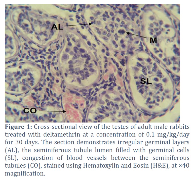
It was observed that most of the seminiferous tubules exhibited disorganized germ cell layers, and their lumen was filled with germ cells, along with occurrence of Sertoli cell vacuolation. Additionally, congestion of blood vessels between the seminiferous tubules was evident, and there was an increase in Leydig cell numbers with reduced distance between adjacent seminiferous tubules in certain histological sections, as illustrated in Figure (1).
Dose-dependent effect of deltamethrin on Leydig cells
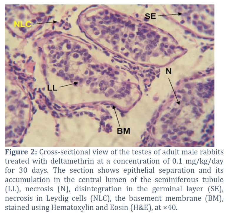
In some other sections, shrinkage of the seminiferous tubule lumen was observed, along with dissociation of the germinal lineage cells and separation of the germinal epithelium from the basement membrane. These cells accumulated in the central lumen of the seminiferous tubule. Additionally, evidence of degeneration and disintegration in the germinal layer and Leydig cells was also noted, as shown in Figure (2).
Dose-dependent effect of deltamethrin on the walls of the seminiferous tubules
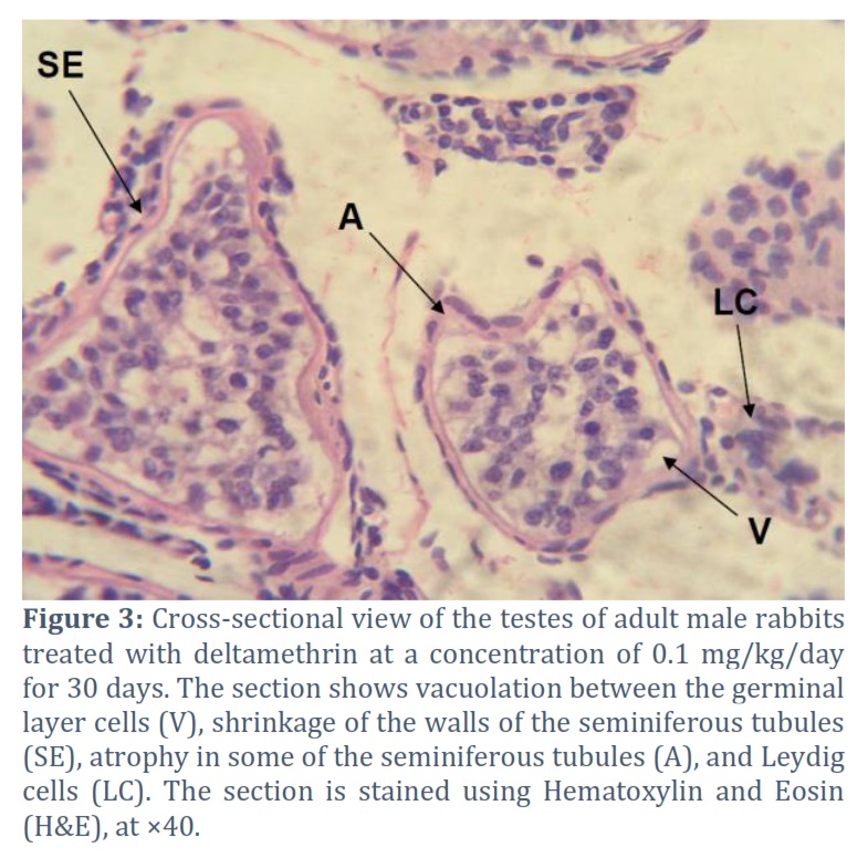
Moreover, the current study revealed significant histological alterations in the seminiferous tubules. These alterations were characterized by irregular and undulating appearance of the tubule walls, as well as atrophy in certain sections of the seminiferous tubules. Additionally, there was irregularity observed in the germinal epithelium, and intriguingly, vacuolation was observed between some regions of the germinal layer, as illustrated in Figure (3).
Dose-dependent effect of deltamethrin on macrophages and inflammatory cells
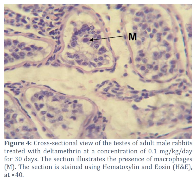
Furthermore, the study demonstrated the presence of large multinucleated cells within the seminiferous lumen, as depicted in Figure (4). Concurrently, notable congestion of blood vessels and infiltration of inflammatory cells near the vascular structures were also observed, as illustrated in Figure (1). These findings indicate the occurrence of inflammation and vascular changes within the testicular tissue.
The effect of increasing the dose of deltamethrin on testicular tissue
In a related context, the histological results of the testes in rabbits treated with 0.3 mg/kg concentration of deltamethrin for a duration of 30 days revealed more pronounced tissue alterations compared to the previous groups.
Dose-dependent effect of deltamethrin on the lumen of seminiferous tubules and the basement membrane
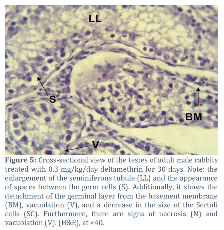
These alterations were characterized by the occurrence of dilation in some of the seminiferous tubules and the presence of small spaces between Sertoli cells and the detachment of the germinal epithelium from the basal membrane. Additionally, vacuolation was observed in certain regions of the seminiferous tubules, as well as the occurrence of vacuoles between adjacent Sertoli cells, as depicted in Figure (5). Furthermore, there was noticeable occurrence of disorganization in the Sertoli cells, leading to an increase in intercellular space between neighboring Sertoli cells, as illustrated in Figure (5). These findings indicate significant disruptions in the testicular tissue structure and function due to exposure to the deltamethrin pesticide at the specified concentration and duration.
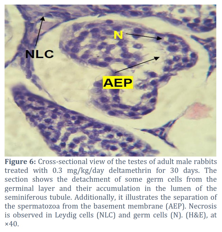
Similarly, observations revealed the detachment of some germinal epithelial cells and their accumulation within the lumen of the seminiferous tubules, leading to the separation of the spermatic epithelium from the basal membrane (Epithelial separation), as depicted in Figure (6).
Dose-dependent effect of deltamethrin on sperm cells
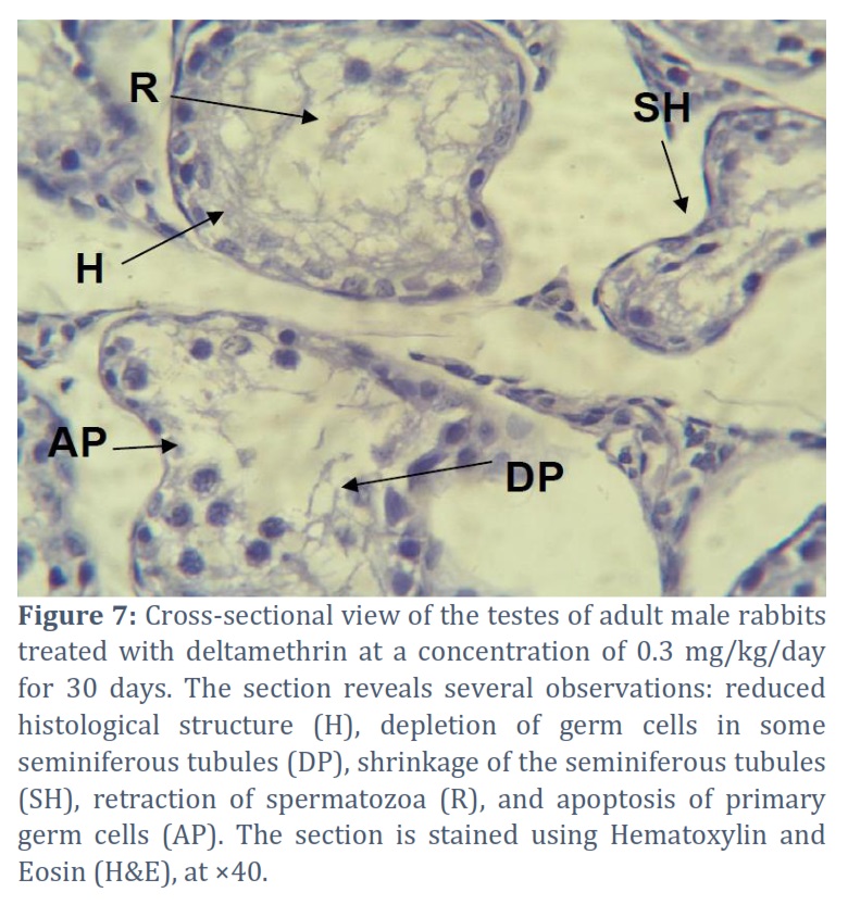
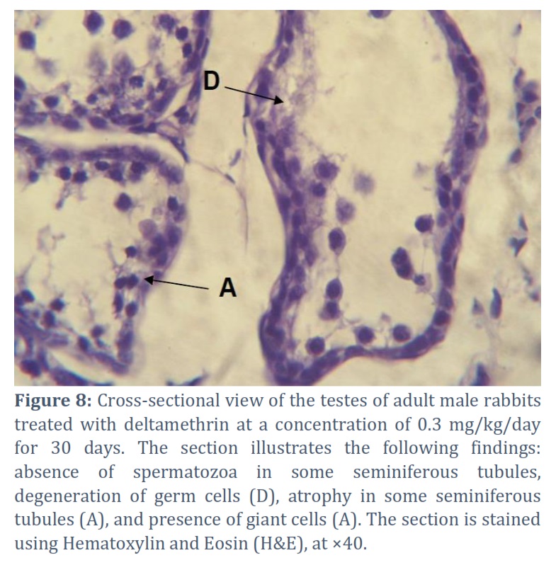
Furthermore, there was an increase in the contraction of the seminiferous tubules and depletion in certain germinal cell layers, resulting in degeneration, vacuolation, and apoptosis of spermatogonia, Primary sperm cells, spermatids, and mature spermatozoa, with the retraction of spermatids and mature spermatozoa back into the seminiferous tubules, as illustrated in Figure (7). On the other hand, the study indicated the absence of spermatozoa in some seminiferous tubule lumens and revealed the presence of Sertoli cells within the lumen of the seminiferous tubules, along with an increase in fibrosis between the seminiferous tubules, as demonstrated in Figure (8). These findings collectively highlight significant disruptions in spermatogenesis and impaired sperm production due to the effects of the deltamethrin pesticide on the testicular tissue.
Figures & Tables
The current study revealed noticeable histological changes in rabbits exposed to concentrations of 0.1 and 0.3 mg/kg of deltamethrin. These changes include alterations in the thickness of the seminiferous tubule walls, dissociation of Sertoli cells, separation of the germinal epithelium from the basal membrane, and their shrinkage. The results of this study are consistent with the findings of Aboelwafa et al., [49] in their research on the preventive effects of melatonin supplementation against taxol-induced testicular cytotoxicity in adult rats. Aboelwafa et al. reported that the basal lamina plays a crucial role in maintaining the transportation of materials between interstitial tissues and the spermatogenic germinal epithelium, thus preserving the structure and function of these tissues. In their study, Ghanami Gashti et al., [50] indicated that the thickness of the seminiferous tubule wall weakens its relationship with the interstitial tissue. Moreover, an increase in the wall thickness leads to the manifestation of several pathological disturbances within the testis, particularly affecting the function of Sertoli cells. These disturbances have an impact on the differentiation of germ cells and inhibit the formation of spermatozoa. In their study on mice, Wang et al., [51] demonstrated that Sertoli cells secrete Collagen fibers IV, which lead to thickening of the walls of the seminiferous tubules. Consequently, this process results in impaired spermatogenesis and a reduction in sperm formation.
The current study also revealed the absence of sperm in some seminiferous tubules. Moreover, the seminiferous tubules exhibited areas of detachment within the interstitial tissue, along with an increase in the distance between germ cells. Additionally, germ cells were found to detach and accumulate within the lumen of the seminiferous tubules. Indeed, these results are consistent with what Mostafa et al., [52] reported in their study on the effects of permethrin, a pesticide, on the testes of adult albino rats. The findings in both studies indicate similar alterations in the seminiferous tubules, such as detachment and accumulation of germ cells, as well as changes in the interstitial tissue, which align with the toxic effects of permethrin on the testicular structure. Ghanami Gashti and others [50] highlighted in their study that the occurrence of a disorder in Sertoli cells will undoubtedly affect germ cells and eventually lead to tissue abnormalities in the testes. These findings are consistent with the results of Wang and others [51] regarding the impact of Sertoli cell syndrome on the testicular histology and genetic mechanisms in male albino mice. Washburn et al. [53] stated in their study that Sertoli cells play a crucial role in the development of germ cells by forming the blood-testis barrier, which protects and nourishes the germ cells and facilitates the transport of nutrients and hormones to them. It is believed that all these pathological signs are due to a dysfunction in the structure and function of Sertoli cells. The results of this study also showed the presence of large phagocytic cells within the lumen of the seminiferous tubules. Researchers have differed in identifying the origin of these cells. Some have suggested that they are true phagocytes, while others have attributed them to lymphoid cells or identified them as Sertoli cells with phagocytic activity, which engulf residual bodies from spermatozoa under normal conditions. The current study revealed that deltamethrin has an effect on Leydig cells. Indeed, the results of the current study are consistent with the findings of Zirkin and Papadopoulos [54] in their research on the immune effects of Sertoli cells. They indicated that Leydig cells serve as a center for regulating fertility by producing the hormone testosterone. They also clarified in their study that Leydig cells are stimulated by luteinizing hormone (LH), which in turn stimulates the production of arachidonic acid and male hormones. The results of the current study revealed that injecting rabbits with a concentration of 0.3 mg/kg of deltamethrin led to an increase in degenerative changes in the epithelium of seminiferous tubules, shrinkage of the spermatic tubules, depletion of certain germinal cell layers in the seminiferous tubules, and programmed cell death in germ cells. Moreover, the study showed a retraction of spermatogonia and mature sperm back into the seminiferous tubules. Additionally, numerous seminiferous tubules were found to be devoid of germ cells, indicating that the effect of the pesticide is dose-dependent. With an increase in the concentration of the dose, the degenerative changes were more pronounced. This effect is believed to be associated with a disruption in Sertoli cells, which, in turn, affects the crucial proteins necessary for the differentiation of germ cells. These proteins are secreted at their highest levels during the differentiation stage of spermatogonia. This result is consistent with what Doyle et al., [55] found in their study regarding the characteristics of sperm and the histological structure of testes in mice after long-term exposure to the compound (Di-(2-ethylhexyl) Phthalate). This result is also consistent with what Moreira et al., [56] mentioned in their study on fertilization and reproductive toxicity mechanisms due to pesticide exposure. They suggested that the retrograde movement of spermatids and mature sperm within the seminiferous tubule wall might be a response to testicular toxicity induced by deltamethrin.
The treatment with deltamethrin at different doses (whether low or high doses) for one month caused severe pathological tissue damage in the testes, characterized by congestion, hemorrhage, vacuolation, and detachment of a portion of the germinal epithelium from the basement membrane.
Conflict of Interest
The authors declare no conflict of interest.
T. A. Ibrahim planned the study, T. A. Ibrahim and F. S. Abd Al-Latif executed the experiment and performed lab work, M. N. Abbas contribute to preparations of chemicals and technical writing of the manuscript.
![]() References
References
- Khaleel LR, Al-Hermizy SM, Abbas MN. Statistical Indicators for Evaluating the Effect of Heavy Metals on Samaraa Drug Industry Water Exposed to the Sun and Freezing. Tropical Journal of Natural Product Research, (2022); 6(12): 1969-1974.
- Ibrahim TA, Abbas MN, Abbas FS. Detoxification of Pesticides Wastewater by Adsorption Technique Feasibility of Agricultural Waste Utilization. (2016); 1-5; LAMBERT Academic Publishing
- Abbas MN, Ali ST, Abbas RS. Rice Husks as a Biosorbent Agent for Pb+2 Ions from Contaminated Aqueous Solutions: A Review. Biochemical and Cellular Archives, (2020); 20(1): 1813-1820.
- Hashem NS, Ali GAA, Jameel HT, Khurshid AN, Abbas MN. Heavy Metals Evaluation by Atomic Spectroscopy, for Different Parts of Water Hyacinth (Eichhornia crassipes) Plants Banks of Tigris River. Biochemical and Cellular Archives, (2021); 21(2): 3813-3819
- Alalwan HA, Mohammed MM., Sultan AJ., Abbas MN, Ibrahim TA, Aljaafari HAS, Alminshid AA. Adsorption of methyl green stain from aqueous solutions using non-conventional adsorbent media: Isothermal kinetic and thermodynamic studies. Bioresource Technology Reports, (2021); 14, Article number: 100680.
- Abbas MN, Alalwan HA. Catalytic Oxidative and Adsorptive Desulfurization of Heavy Naphtha Fraction. Korean Journal of Chemical Engineering, (2019); 12(2): 283-288.
- Alalwan HA, Abbas MN, Alminshid AH. Uptake of Cyanide Compounds from Aqueous Solutions by Lemon Peel with Utilising the Residue Absorbents as Rodenticide. Indian Chemical Engineer, (2020); 62(1): 40-51
- Ibrahim SA, Hasan MB, Al-Tameemi IM, Ibrahim TA, Abbas MN. Optimization of adsorption unit parameter of hardness remediation from wastewater using low-cost media. Innovative Infrastructure Solutions, (2021); 6(4) Article number: 200
- Abbas MN. Phosphorus removal from wastewater using rice husk and subsequent utilization of the waste residue. Desalination and Water Treatment, (2015); 55(4): 970-977.
- Abbas MN, Abbas FS. Application of Rice Husk to Remove Humic Acid from Aqueous Solutions and Profiting from Waste Leftover. WSEAS Transactions on Biology and Biomedicine, (2014); 11:62-69.
- Abbas MN, Abbas FS. Iraqi Rice Husk Potency to Eliminate Toxic Metals from Aqueous Solutions and Utilization from Process Residues. Advances in Environmental Biology, (2013); 7(2): 308-319
- Abbas MN, Al-Madhhachi AT, Esmael SA. Quantifying soil erodibility parameters due to wastewater chemicals. International Journal of Hydrology Science and Technology, (2019); 9(5): 550-568
- Ali GAA, Ibrahim SA, Abbas MN. Catalytic Adsorptive of Nickel Metal from Iraqi Crude Oil using non-Conventional Catalysts. Innovative Infrastructure Solutions, (2021); 6(7): 1-9
- Abbas MN, Abbas FS. The Predisposition of Iraqi Rice Husk to Remove Heavy Metals from Aqueous Solutions and Capitalized from Waste Residue. Research Journal of Applied Sciences, Engineering and Technology, (2013); 6(22): 4237-4246
- Maddodi SA, Alalwan HA, Alminshid AH, Abbas MN. Isotherm and computational fluid dynamics analysis of nickel ion adsorption from aqueous solution using activated carbon. South African Journal of Chemical Engineering, (2020); 32: 5-12
- Abbas MN, Abbas FS. The Feasibility of Rice Husk to Remove Minerals from Water by Adsorption and Avail from Wastes. Research Journal of Applied Sciences, WSEAS Transactions on Environment and Development, (2013); 9(4): 301-313
- Alalwan HA, Abbas MN, Abudi ZN, Alminshid AH. Adsorption of thallium ion (Tl+3) from aqueous solutions by rice husk in a fixed-bed column: Experiment and prediction of breakthrough curves. Environmental Technology and Innovation, (2018); 12: 1-13
- Abbas MN, Nussrat TH. Statistical Analysis of Experimental Data for Adsorption Process of Cadmium by Watermelon Rinds in Continuous Packed Bed Column. International Journal of Innovation, Creativity and Change, (2020); 13(3): 124-138.
- Abdullah WR, Alhamadani YAJ, Abass IK, Abbas MN. Study of chemical and physical parameters affected on purification of water from inorganic contaminants. Periodicals of Engineering and Natural Sciences, (2023); 11(2): 166-175
- Al-Ali SIS, Ibrahim SA, Abbas MN, Ibrahim TA. Mathematical Modelling of Selenium Adsorption using Pomegranate Peel. Tikrit Journal of Engineering Sciences, (2023); under publishing.
- Hasan MB, Al-Tameemi IM, Abbas MN. Orange Peels as a Sustainable Material for Treating Water Polluted with Antimony. Journal of Ecological Engineering, (2021); 22(2): 25-35
- Al-Hermizy SMM, Al-Ali SIS, Abdulwahab IA, Abbas MN. Elimination of Zinc Ions (Zn+2) from Synthetic Wastewater Using Lemon Peels. Asian Journal of Water, Environment and Pollution, (2022); 19(5): 79-85
- Al-Ali SIS., Abudi ZN, Abbas MN. Modelling and Simulation for the use of Natural Waste to Purified Contaminated Heavy Metals. Journal of the Nigerian Society of Physical Sciences, (2023); 5(1): Article No.: 1143
- Ali SAK, Almhana NM, Hussein AA, Abbas MN. Purification of Aqueous Solutions from Toxic Metals using Laboratory Batch Mode Adsorption Unit Antimony (V) Ions as a Case Study. Journal of Green Engineering (JGE), (2020); 10(11): 10662-10680.
- Abbas MN, Al-Hermizy SMM, Abudi ZN, Ibrahim TA. Phenol Biosorption from Polluted Aqueous Solutions by Ulva lactuca Alga using Batch Mode Unit. Journal of Ecological Engineering, (2019); 20(6): 225–235
- Ali GAA, Abbas MN. Atomic Spectroscopy Technique Employed to Detect the Heavy Metals from Iraqi Waterbodies Using Natural Bio-Filter (Eichhornia crassipes) Thera Dejla as a Case Study. Systematic Reviews in Pharmacy, (2020); 11(9): 264-271
- Abudi ZN, Hu Z, Alkhafaji RA. Anaerobic co-digestion of mango leaves and pig manure: performance assessment and kinetic analysis. Biomass conversion and biorefinery, (2022); 12(2): 275-285
- Ghulam NA, Abbas MN, Sachit DE. Preparation of synthetic alumina from aluminium foil waste and investigation of its performance in the removal of RG-19 dye from its aqueous solution. Indian Chemical Engineer, (2020); 62(3): 301-313
- Abbas MN, Ibrahim SA. Catalytic and thermal desulfurization of light naphtha fraction. Journal of King Saud University – Engineering Sciences, (2020); 32(4): 229-235
- Abbas MN, Al-Tameemi IM, Hasan MB, Al-Madhhachi AT. Chemical Removal of Cobalt and Lithium in Contaminated Soils using Promoted White Eggshells with Different Catalysts; South African Journal of Chemical Engineering, (2021); 35: 23-32
- Abbas FS, Abdulkareem WS, Abbas MN. Strength Development of Plain Concrete Slabs by the Sustainability Potential of Lead-Loaded Rice Husk (LLRH). Journal of Applied Engineering Science, (2022); 20(1): 160-167
- Abdulkareem WS, Aljumaily HSM, Mushatat HA, Abbas MN. Management of Agro-Waste by Using as an Additive to Concrete and Its Role in Reducing Cost Production: Impact of Compressive Strength as a Case Study. International Journal on “Technical and Physical Problems of Engineering” (IJTPE), (2023); 54, 15(1): 62-67.
- Abd ali IK, Ibrahim TA, Farhan AD, Abbas MN, Study of the effect of pesticide 2,4-D on the histological structure of the lungs in the albino mice (Mus musculus). Journal of Pharmaceutical Science and Research, (2018); 10(6): 1418-1421
- Abbas MN, Ibrahim SA, Abbas ZN, Ibrahim TA. Eggshells as a Sustainable Source for Acetone Production. Journal of King Saud University – Engineering Sciences, (2021); 34(6): 381-387
- Alminshid AH, Abbas MN, Alalwan HA, Sultan AJ, Kadhome MA. Aldol condensation reaction of acetone on MgO nanoparticles surface: An in-situ drift investigation. Molecular Catalysis, 501, Article No.: 111333
- Alwan EK, Hammoudi AM, Abd IK, Abd Alaa MO, Abbas MN. Synthesis of Cobalt Iron Oxide Doped by Chromium Using Sol-Gel Method and Application to Remove Malachite Green Dye. NeuroQuantology, (2021); 19(8): 32-41
- Abdel-Daim MM, Abuzead SM, Halawa SM, Protective role of Spirulina platensis against acute deltamethrin-induced toxicity in rats. PloS one, (2013); 8(9), e72991
- Gündüz E, Ülger BV, İbiloğlu İ, Ekinci A, Dursun R, et al. Glutamine provides effective protection against deltamethrin-induced acute hepatotoxicity in rats but not against nephrotoxicity. Medical science monitor: international medical journal of experimental and clinical research, (2015); 21: 1107–1114
- Li S, Zheng X, Zhang X, Yu H, Han B, et al. Exploring the liver fibrosis induced by deltamethrin exposure in quails and elucidating the protective mechanism of resveratrol. Ecotoxicology and Environmental Safety, (2021); 207: 111501
- Ilyas WM., Chavan G, Gadkari C, Deltamethrin Poisoning Mimicking Organophosphate Poisoning: A Case Report. Cureus, (2023); 15(1): e34303
- Farrag A- RH, Shalby SEM. Comparative Histopathological and Histochemical Studies on IGR, Lufenuron and Profenofos Insecticide Albino Rats. Journal of Applied Sciences Research, (2007); 3(5): 377-386.
- Ibrahim TA, Mahdi HS, Abbas RS, Abbas MN, Study the Effect of Ribavirin Drug on the histological structure of the testes in Albino mice (Mus musculus). Journal of Global Pharma Technology, (2020); 12(02 Suppl.): 142-146.
- Ibrahim TA, Mohammed AM, Abd ali IK, Abbas MN, Hussien SA. Teratogenic Effect of Carbamazepine Drug on the Histological Structure of Testes in the Albino Mouse (Mus musculus). Indian Journal of Forensic Medicine & Toxicology, (2020); 14(4): 1829-1834
- Kumar A, Sasmal D, Sharma N, Mechanism of deltamethrin induced thymic and splenic toxicity in mice and its protection by piperine and curcumin: in vivo study. Drug and chemical toxicology, (2018); 41(1): 33–41
- Pitzer EM, Sugimoto C, Regan SL, Gudelsky GA, Williams MT, Vorhees CV, Developmental deltamethrin: Sex-specific hippocampal effects in Sprague Dawley rats. Current research in toxicology, (2022); 3, Article No.: 100093
- Mohammed N, Hassan H, Ali A, Khaled F, Mohamed S. Histopathological Alterations in Liver of Male Rabbits Exposed to Deltamethrin and the ameliorative Effect of Folic Acid. AlQalam Journal of Medical and Applied Sciences (AJMAS), (2022); 5(2): 454-460
- Bouzar AC, Benali Y, Bitam A. A low-dose protective effect of phycocyanin on the toxicity of deltamethrin to vital organs in rats: in vivo study. Journal of Fundamental and Applied Sciences, (2020); 12(1): 149-166
- Ali ST, Qadir HT, Moufak SK, Al-Badri MAM, Abbas MN. A Statistical Study to Determine the Factors of Vitamin D Deficiency in Men the City of Baghdad as a Model. Indian Journal of Forensic Medicine & Toxicology, (2020); 14(1): 691-696.
- Aboelwafa HR, Ramadan RA, El-Kott AF, Abdelhamid FM. The protective effect of melatonin supplementation against taxol-induced testicular cytotoxicity in adult rats. Brazilian journal of medical and biological research = Revista brasileira de pesquisas medicas e biologicas, (2022); 55, Article No. e11614
- Ghanami Gashti N, Sadighi Gilani MA, Abbasi M. Sertoli cell-only syndrome: etiology and clinical management. Journal of assisted reproduction and genetics, (2021); 38(3), 559–572
- Wang HX, Zhang R, Li Z, Wang LS., Yu Y, et al. Cypermethrin induces Sertoli cell apoptosis through mitochondrial pathway associated with calcium. Toxicology research, (2021); 10(4), 742–750
- Mostafa HS, Abd El-Baset SA, Kattaia AA, Zidan RA, Al Sadek MM. Efficacy of naringenin against permethrin-induced testicular toxicity in rats. International journal of experimental pathology, (2016); 97(1); 37–49
- Washburn RL, Hibler T, Kaur G, Dufour JM. Sertoli Cell Immune Regulation: A Double-Edged Sword. Frontiers in Immunology, (2022); 13: 913502
- Zirkin BR, Papadopoulos V. Leydig cells: formation, function, and regulation. Biology of reproduction, (2018); 99(1), 101–111
- Doyle TJ, Bowman JL, Windell VL, McLean DJ, Kim KH. Transgenerational Effects of Di-(2-ethylhexyl) Phthalate on Testicular Germ Cell Associations and Spermatogonial Stem Cells in Mice, Biology of Reproduction, (2013); 88(5): 112, 1–15
- Moreira S, Pereira SC, Seco-Rovira V, Oliveira PF, Alves MG, Pereira ML. Pesticides and Male Fertility: A Dangerous Crosstalk. Metabolites, (2021); 11(12); 799.
This work is licensed under a Creative Commons Attribution-Non Commercial 4.0 International License. To read the copy of this license please visit: https://creativecommons.org/licenses/by-nc/4.0


