

Full Length Research Article
Prevalence and Antibiotic Sensitivity Testing of Bacteria Isolated from Open Wounds of Hospitalized Patients
Iqra Arooj1 ⃰, Maryam Noor 1, Nadia Jabbar1, Asghar Javaid2
Adv. life sci., vol. 10, no. 2, pp. 180-185, June 2023
*– Corresponding Author: Iqra Arooj Sulieman (iqra.6051@wum.edu.pk)
Authors' Affiliations
2. Pathology Department, Nishtar Medical University, Multan – Pakistan
[Date Received: 05/09/2022; Date Revised: 14/02/2023; Date Published Online: 30/06/2023; Date Updated:11/09/2025]
Editorial Expression of Concern
20 June 2025: Following publication of this paper, the internal audit (consequent to concerns on quality raised by Web of Science) notified Advancements in Life Sciences about errors in metadata. By this Editorial Expression of Concern, we alert the scientific community of this incidence as we do the required corrections.
Editorial Note
24 June 2025: You are viewing the latest version of this article having correction in article info (metadata) section. Expression of concern is hereby revoked.
Abstract![]()
Introduction
Methods
Results
Discussion
References
Abstract
Background: Substantial infectivity of open wounds by microorganisms not only precipitates in terms of complexity, morbidity and mortality because of encouraging other fatal comorbidities but also becomes challenging to handle by medical practitioners.
Methods: To ascertain the bioburden associated particularly with bacteria in open wounds, this research work was conducted at the Microbiology and Molecular Genetics department of Women University, Multan in association with Pathology Department of Nishtar Hospital and College, Multan from January to June 2018. 65 different wound samples were collected from different wards and analyzed by highly standard procedures for bacterial isolation and characterization, employing biochemical tests including catalase, oxidase, coagulase, motility, triple-sugar iron (TSI), citrate and indole tests. The isolated bacterial strains were also evaluated for sensitivity or resistance against twelve different antibiotics.
Results: All the 65 samples were found to be positive for bacterial presence. Gram-positive cocci constituted 63% of the isolates and gram-negative rods comprised 37%. Biochemical tests revealed that the predominant pathogen was Staphylococcus aureus (63.1%) followed by Proteus spp., (15.4%), Pseudomonas aeruginosa (12.3%) and Escherichia coli (9.2%), respectively. Antibiotic sensitivity testing disclosed that the most effective antibiotics against these isolates were Tigecycline and Polymyxin B while the least effective antibiotics were Ceftazidime and Ampicillin.
Conclusion: These findings can prove beneficial in understanding the prevalence of various bacteria in wound infections. In addition, they indicate the need to develop and implement antibiotic stewardship programs so as to combat drug resistance among pathogens.
Keywords: Gram-positive cocci; Drug resistance; Staphylococcus aureus
Introduction![]()
Open wound infections are not only emerging as era-based adversity but also becoming a matter of concern for medical practitioners and researchers across the globe as a consequence of sufficient reasons including low consideration towards their microbiological analysis, lack of deep examination of their causes and effects as well as lack of awareness [1, 2]. Open wounds are of many types depending on size, complexity, pus formation, location or site and severity of symptoms, among other medical aspects [3, 4].
If the open wounds remain untreated for a long period of time, they turn into chronic wounds (wounds not undergoing the normal cycle of healing) which become difficult to handle and may progress towards further complications and health hazard circumstances [5]. In terms of the relationship between wounds and their demolition rate, diversity of microbial flora and their mass load are considered as the main contributors [6, 7]. There are many possible routes, through which microbes can directly intact with wounds at tissue or cellular level, primarily the natural normal flora located on outermost or inner layers of our main sensing organ, skin, can become opportunistic and proliferate for rooting inside the wound to establish their colonization there and make the state of wound worse. The correlation between wound and microbiota does not only characterize complexity of wound infection but also predict its potential influence on healing [8].
When the log phase of bacterial population reaches its maximum level, it not only endorses the obstruction in healing process but also enhances the possibility of initiation of comorbidities [9]. In terms of wounds resulting from burn injuries, not only the not only from surroundings but also from the contaminated dressings which can become the foremost reason for wound infection [10- 12]. The cost of products pertaining to wound safety and care (such as dressings) is escalating constantly around the globe per annum and are predicted to reach higher rates in the upcoming 4 to 5 years [13, 5].
Wounds with the traces of creamy discharged fluid (dead bodies of white blood cells in bulk amount called as pus) likely indicate that wound has been infected by some living entity (with high chances of bacterial bioburden) either from exogenous environment or some other source, as pus formation is an induced response of immune system against living organisms for their eradication [14- 16]. Among all of the microorganisms causing infection in wounds, Staphylococcus aureus, a medically established pathogenic microbial entity belonging to gram positive species plays a key role in progressive deterioration of wound infection [17, 18]. S. aureus is generally recognized as a highly adaptable opportunistic pathogen in hospitals associated with high degree of infectivity in open wounds, hospital acquired infections, as well as catheterized patients of respiratory or urinary tract infections [19, 20].
Pseudomonas aeruginosa, a well-known member of gram-negative species is recognized for its ability to promote the infection in different categories of wounds such as hospital-acquired wounds, wounds resulting from burning flames or damp natured wounds. It possesses multiple attributes in term of resistivity against a broad range of antibiotics and quick deterioration of cells and tissues leading to sepsis at elevated pace [21, 22]. Escherichia coli as facultative anaerobic coliform bacterium and Proteus species belonging to gram negative genera have also been reported to induce infections in wounds particularly in wounds of patients suffering from diabetes mellitus [23, 24]. In a recent study conducted in Pakistan, Staphylococcus aureus was reported as the most prevalent pathogen in burn wounds, followed by Staphylococcus epidermidis, E. coli, Proteus spp., K. pneumoniae and P. aeruginosa [25].
Having adequate information regarding causative microbial agents involved in the course of wound infection can potentially facilitate the cure, prevention and control of such infections. Therefore, the present study was designed to identify the diversity and prevalence of common bacteria in open wounds of patients as well as to ascertain their antibiotic susceptibility profile. It was anticipated that the knowledge regarding antibiotic resistance patterns among common pathogens will prove helpful in formulating efficient therapeutic plan to treat wound infections.
Methods![]()
Sample collection and ethical considerations
The present research work was conducted at the Microbiology and Molecular Genetics department of The Women University, Multan in collaboration with Pathology Department of Nishtar Hospital and College, Multan from January to June 2018. 65 wound samples from different wards including the burn unit of Nishtar Hospital were collected strictly following the rules of Institutional Research Ethics Committee to avoid any controversy regarding personal consent and ethical beliefs of patients. All the data collected from patients was held confidential. Informed consent was obtained from patients or their relatives. The samples were collected randomly with respect to gender, age and disease or symptoms. Samples were taken when the wounds of patients were in unwashed condition without any medication due to greater chances of bacterial presence on wound areas under such conditions. Sampling was done using sterilized swab sticks subsequent to which samples were brought to the Microbiology laboratory and processed within 2 to 3 hours of sample collection.
Bacterial isolation and phenotypic characterizations
The culturing techniques considered as standard methods for the purpose of isolating bacteria were employed in the laboratory under the recommended sterile conditions. For culturing fastidious microbiome blood agar, an enriched medium and MacConkey agar, a differential medium was used. Culture plates were prepared as per prescribed recipes by maintaining contamination free conditions [26]. The samples were immediately inoculated onto the surface of prepared petri dishes using the sample swabs and then incubated at 37°C under aerobic atmospheric environment. After completion of 24 hours incubation period, the obtained bacterial colonies were morphologically characterized based on their appearance, texture etc. by keen visual observation. Gram staining was employed as standard method for the purpose of viewing detailed cell morphology of obtained bacterial isolates. For further identification and characterization of isolated bacteria, biochemical testing was performed which comprised of catalase, oxidase, coagulase, motility, triple-sugar iron (TSI), citrate and indole tests.
Antibiotic susceptibility profiling
Obtained bacterial isolates were evaluated for sensitivity or resistance against different selected antibiotics on Mueller Hinton Agar plates pursuing disc diffusion method by means of inoculating 24 hours bacterial cultures using sterile conditions to ensure confluent growth of gram negative as well as gram positive bacteria. Antibiotic discs used for this purpose included Amikacin (AMK), Ampicillin (AMP), Aztreonam (ATM), Ceftazidime (CAZ), Ceftriaxone (CTX), Ciprofloxacin (CIP), Colistin (CST), Imipenem (IPM), Linezolid (LZD), Polymyxin B (PMB), Piperacillin/Tazobactam (TZP) and Tigecycline (TGC). These were aseptically placed onto the surface of agar plates at well-spaced intervals and gently touched with sterile spatula to ensure their firm contact with agar surface. Plates were then incubated at 37°C for 24 hours under sterile conditions. After the completion of incubation period, plates were observed for presence or absence of zones of inhibition around the antibiotic discs. Zones were measured in millimeters using Inhibition Zone Ruler provided by the manufacturer and then categorized in accordance with the standardized chart for antibiotics (M100-S23) provided by Clinical and Laboratory Standards Institute (CLSI).
Results![]()
Colony characteristics and gram staining
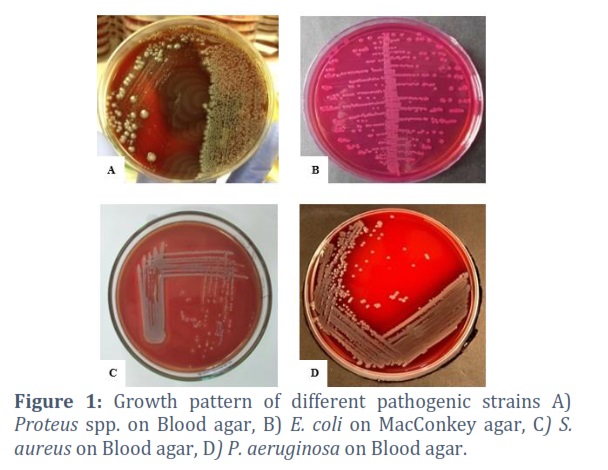
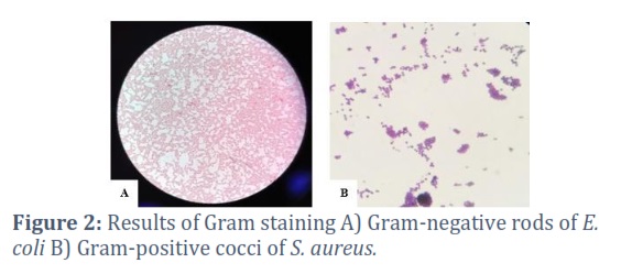
All the 65 samples of wounds collected from patients were positive for bacterial pathogens. One isolate from each sample was selected for visual morphological examination and biochemical characterization. According to keen observation, it was revealed that all the 65 isolates were diverse in terms of colony characteristics including size, appearance, elevation, margin etc. indicating that individual colonies were dissimilar to each other (Fig 1). Gram staining procedure was employed for obtaining cell morphological details of obtained isolates and it was revealed that the samples comprised of both gram-positive and gram-negative bacteria (Fig 2). Sixty three percent (N=41) isolates were gram positive cocci, and thirty seven percent (N=24) were gram negative rods.
Biochemical characterization of isolates
Biochemical tests were followed in a series for the further characterization of bacterial isolates. Coagulase and catalase test were performed for gram positive bacteria whereas for gram negative bacteria catalase, coagulase, indole, TSI, oxidase, motility and citrate tests were performed. Sequential completion of biochemical procedure indicated that some bacteria were positive while others were negative for corresponding tests. Results indicated that all (N=65) the gram-positive and gram-negative bacterial strains were positive for catalase test. 63% (N=41) of the isolated bacteria were positive for coagulase test while 37% (N=24) were negative. All (N=24) the gram-negative bacterial strains were indicated as positive for motility test. For the citrate test, 75% (N=18) produced positive result while rest of 25% (N=6) were negative for this test. 33% (N=8) were found to be positive for oxidase test while 67% (N=16) were negative. For the indole test, 25% (N=6) produced positive result while rest of 75% (N=18) were negative for this test. For TSI test, 42% (N=10) produced acid in butt only along with gas production, 33% (N=8) did not produce acid or gas and the remaining 25% (N=6) produced acid as well as gas. The isolated gram-positive bacterial species from wounds were majorly consisting of Staphylococcus aureus that represented 63.1% (N=41) of total isolates. The gram-negative bacterial isolates 15.4% (N=10) of Proteus species, 12.3% (N=8) of Pseudomonas aeruginosa and 9.2% (N=6) of Escherichia coli.
Antibiotic sensitivity pattern of isolates
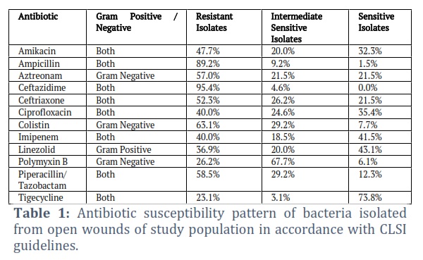
The isolated strains including gram-positive as well as gram-negative bacteria were evaluated for antibiotic sensitivity against twelve different selected antibiotics, and it was observed that all of the bacterial strains with the exception of one showed resistance against three or more than three different antibiotics and hence could be referred to as multidrug resistant organisms. The antibiotic susceptibility pattern outlined in Table 1 indicates that majority of the isolated bacterial strains were resistant to Ampicillin (AMP), Aztreonam (ATM), Ceftazidime (CAZ), Ceftriaxone (CTX), Colistin (CST) and Piperacillin/Tazobactam (TZP) with the number of resistant isolates being 58, 37, 62, 34, 41 and 38, respectively. Furthermore, majority of the bacterial isolated were either sensitive or intermediate sensitive to Amikacin (AMK), Ciprofloxacin (CIP), Imipenem (IPM), Linezolid (LZD), Polymyxin B (PMB) and Tigecycline (TGC), the number of sensitive or intermediate sensitive isolates being 34, 39, 39, 41, 48 and 50, respectively.
Figures & Tables
Wound is said to be characterized by any sort of damage to skin tissue and it provides the chance to billions of microorganisms residing in the surrounding atmosphere to gain entrance into the wound and produce their progeny to reach at maximum possible level so as to support infectious state of wound. Many microbes are directly correlated with widespread wounds infections and to achieve sufficient knowledge about them is mandatory for curing and controlling such infections. The present research was carried out to analyze the bacterial load in samples of open wounds collected from different wards of Nishtar Hospital, Multan including Burn Unit and the isolates were also evaluated for susceptibility against selected antibiotics. Results of present research work revealed that all the samples were infected with one or more than one type of bacteria and comprised of both gram positive as well as gram negative bacterial flora. The percentage of gram-positive isolates was high as compared to gram-negative strains. The present results were in accordance with findings of Maharjan et al in 2020 and Regmi et al in 2020 [27, 28]. According to the conclusion of their research work, the wounds get infected primarily by gram-positive isolates (70.4% and 60.6%, respectively) as compared to gram negative isolates (29.6% and 38.6%, respectively). On the contrary, another study reported that 23.56% of the isolates were gram positive and 76.44% were gram negative from the pus or discharged fluid of wounds [16]. These differences could be attributed towards the differences in population from which samples were collected as microbial prevalence varies considerably depending on geographical origin.
On the basis of biochemical testing, it was evaluated that the isolated bacterial species obtained from wounds of patients were primarily consisting of gram-positive Staphylococcus aureus (63%) followed by gram-negative bacterial isolates including Proteus spp. (15%), Pseudomonas aeruginosa (12%) and Escherichia coli (9%), respectively. Previously, it was described that Staphylococcus aureus is directly involved in causing infection in different tissue and bone wounds as they found the prevalence of this causative bacterium to be 51.8% in wounds [29]. Staphylococcus aureus as being responsible for causing wound infections especially in hospital environment has been evidently proved by recent researches also [27, 28]. Ali et al in 2017 isolated Staphylococcus aureus from patients of urinary tract infections by following the same techniques and procedures as used in present research work [30]. In another study, Escherichia coli and Pseudomonas aeruginosa were reported for dominantly causing infectivity in wounds with pus discharge [16]. Earlier, it has also been reported that Pseudomonas aeruginosa exhibits significant positive correlation with wound infections [22].
Based on antibiotic sensitivity profiling, it was observed that Tigecycline and Polymyxin B were the most effective antibiotics against isolated bacteria whereas, Ceftazidime and Ampicillin were the least effective. In another study, of all the 9 classes of antibiotics tested, Augmentin and Ciprofloxacin were found to be the most effective against reasonable percentage of bacterial isolates, while polymyxin B and Amoxycillin were found to be least effective. This was attributed towards indiscriminate and empirical use of these drugs [31]. Mu’azu et al in 2021 conducted similar work for the characterization and identification of Staphylococcus aureus from different wounds infections (such as wounds created by bite etc) and found out predominant resistance against Methicillin [20].
The study was limited to the antibiotic resistance profiling of pathogens by disc-diffusion method and the underlying genetic basis of drug resistance was not studied. In addition, sample size was limited for the present study and in future, research employing substantial population size and focusing on genetic mechanisms may contribute more reliable insights into the ever-evolving antimicrobial susceptibility pattern of bacteria.
Open wounds should be treated as soon as possible because their exposure to environment easily causes infections by natural flora of human skin or thousands of microbes roaming in air and for adequate treatment, periodic surveillance of antimicrobial sensitivity profile is mandatory. Well-recognized pathogenic strains such as Staphylococcus aureus, Escherichia coli, Proteus spp. and Pseudomonas aeruginosa must be treated with great concern as they are becoming resistant towards more and more antibiotics with the passage of time.
Conflict of Interest
The authors declare that there is no conflict of interest.
Performed the experiments: Maryam Noor, Nadia Jabbar
Analyzed the data: Maryam Noor, Iqra Arooj, Nadia Jabbar
Contributed materials/ analysis/ tools: Iqra Arooj, Asghar Javaid
Wrote the paper: Maryam Noor, Iqra Arooj, Nadia Jabbar, Asghar Javaid
![]()
References
- Li J, Wang Q, Lu Y, Feng Q, He X, Li MDZ, Zhang K. Relationship between time to surgical debridement and the incidence of infection in patients with open tibial fractures. Orthopaedic Surgery, (2020);12(2): 524-532.
- Monnheimer M, Cooper P, Amegbletor HK, Pellio T, Groß U, Pfeifer Y, Schulze MH. High prevalence of carbapenemase-producing Acinetobacter baumannii in wound infections, Ghana, 2017/2018. Microorganisms, (2021); 9(3): 537.
- Xu Z, Han S, Gu Z, Wu J. Advances and impact of antioxidant hydrogel in chronic wound healing. Advanced Healthcare Materials, (2020); 9(5): 1901502.
- Li S, Renick P, Senkowsky J, Nair A, Tang L. Diagnostics for wound infections. Advances in Wound Care, (2021); 10(6): 317-327.
- Sen CK. Human wounds and its burden: An updated compendium of estimates. Advances in Wound Care, (2019); 8(2):39–48.
- Loesche M, Gardner SE, Kalan L, Horwinski J, Zheng Q, Hodkinson BP, … Grice EA. Temporal stability in chronic wound microbiota is associated with poor healing. Journal of Investigative Dermatology, (2017); 137(1): 237-244.
- Haidari H, Kopecki Z, Sutton AT, Garg S, Cowin AJ, Vasilev K. pH-responsive “smart” hydrogel for controlled delivery of silver nanoparticles to infected wounds. Antibiotics, (2021); 10(1): 49.
- Norman G, Shi C, Westby MJ, Price BL, McBain AJ, Dumville JC, Cullum N. Bacteria and bioburden and healing in complex wounds: A prognostic systematic review. Wound Repair and Regeneration, (2021);29(3): 466-477.
- Le L, Baer M, Briggs P, Bullock N, Cole W, DiMarco D, … Serena TE. Diagnostic accuracy of point-of-care fluorescence imaging for the detection of bacterial burden in wounds: results from the 350-patient fluorescence imaging assessment and guidance trial. Advances in Wound Care, (2021); 10(3): 123-136.
- Nedomansky J, Maier B, Rath T, Radtke C. Current challenges in the treatment of paediatric burn patients – a retrospective experience at a Viennese burn unit. HandchirurgieMikrochirurgiePlastischeChirurgie, (2019); 51(02): 94-101.
- Abdalla SSI, Katas H, Chan JY, Ganasan P, Azmi F, Fauzi MB. Gelatin hydrogels loaded with lactoferrin-functionalized bio-nanosilver as a potential antibacterial and anti-biofilm dressing for infected wounds: synthesis, characterization, and deciphering of cytotoxicity. Molecular Pharmaceutics, (2021); 18(5): 1956-1969.
- El-Ghoul Y, Alminderej FM. Bioactive and superabsorbent cellulosicdressing grafted alginate and Carthamus tinctorius polysaccharide extract for the treatment of chronic wounds. Textile Research Journal, (2021); 91(3-4): 235-248.
- Nussbaum SR, Carter MJ, Fife CE, DaVanzo J, Haught R, Nusgart M, Cartwright D. An economic evaluation of the impact, cost, and medicare policy implications of chronic nonhealing wounds. Value Health, (2018); 21(1): 27-32.
- Khan RA, Jawaid M, Khaleel M. Bacteriological profile and antibiogram of isolates from pus samples in a tertiary care centre. International Journal of Current Microbiology and Applied Sciences, (2018); 7(1): 387-394.
- Wadekar MD, Sathish JV, Pooja C. Bacteriological profile of pus samples and their antibiotic susceptibility pattern. Indian Journal of Microbiology Research, (2020); 7(1): 43-47.
- Sharma R, Batra S, Balothia V, Agarwal S. Bacteriological profile and antimicrobial susceptibility pattern of pus culture isolates from a tertiary care hospital, SMS Medical College Jaipur. European Journal of Molecular & Clinical Medicine, (2021); 7(11): 7502-7508.
- Algburi A, Al-Hasani HM, Ismael TK, Abdelhameed A, Weeks R, Ermakov AM, Chikindas ML. Antimicrobial activity of Bacillus subtilis KATMIRA1933 and Bacillus amyloliquefaciens B-1895 against Staphylococcus aureus biofilms isolated from wound infection. Probiotics and Antimicrobial Proteins, (2021); 13(1): 125-134.
- Rowe SE, Beam JE, Conlon BP. Recalcitrant Staphylococcus aureus infections: obstacles and solutions. Infection and Immunity, (2021); 89(4): e00694-20.
- Farahpour MR, Pirkhezr E, Ashrafian A, Sonboli A. Accelerated healing by topical administration of Salvia officinalis essential oil on Pseudomonas aeruginosa and Staphylococcus aureus infected wound model. Biomedicine & Pharmacotherapy, (2020); 128: 110120.
- Mu’azu L, Ali M, Ahmad AM, Zungum IU, Abdallah MS. Isolation, characterization and determination of prevalence rate of methicilin resistance Staphylococcus aureus (MRSA) from different types of wound. Gulf Journal of Molecular Biology, (2021); 1(1): 1-5.
- Vanderwoude J, Fleming D, Azimi S, Trivedi U, Rumbaugh KP, Diggle SP. The evolution of virulence in Pseudomonas aeruginosa during chronic wound infection. Proceedings of the Royal Society B Biological Sciences, (2020); 287(1937): 20202272.
- Raizman R, Little W, Smith AC. Rapid diagnosis of Pseudomonas aeruginosa in wounds with point-of-care fluorescence imaging. Diagnosis, (2021); 11(2): 280.
- Moglad EH, Boon SKA, Ali HT. Various medicinal plants: a promising treatment for multidrug-resistant bacteria isolated from wound infection. International Journal of Pharmaceutical Sciences and Research, (2020); 11(2): 839-843.
- Thabit AG, El-Sabour A, Nafie AMA, El-Mokhtar MA, Biomy YE. Detection of Proteus species in diabetic wounds and their antibiotic resistance profile analysis. Bulletin of Pharmaceutical Sciences,(2020);43(1): 1-10.
- Hubab M, Maab H, Hayat A, Ur Rehman M. Burn wound microbiology and the antibiotic susceptibility patterns of bacterial isolates in three burn units of Abbottabad, Pakistan. Journal of Burn Care & Research. (2020);41(6):1207-1211.
- Cappuccino JG, Sherman N. Microbiology: a laboratory manual. 2014. 10th ed.Harlow, England.
- Maharjan N, Mahawal BS. Bacteriological profile of wound infection and antibiotic susceptibility pattern of various isolates in a tertiary care center. Journal of Lumbini Medical College, (2020); 8(2): 218-224.
- Regmi SM, Sharma BK, Lamichhane PP, Gautam G, Pradhan S, Kuwar R. Bacteriological profile and antimicrobial susceptibility patterns of wound infections among adult patients attending Gandaki Medical College Teaching Hospital, Nepal. Journal of Gandaki Medical College-Nepal, (2020); 13(1): 60-64.
- Samad A, Asghar M, Naeem M, Rehman N, Asghar N, Haroon M. Identification of pathogenic bacteria isolated from tissues, bones infections and their antibiotic susceptibility pattern at Khyber Teaching Hospital, Peshawar. Pakistan Journal of Surgery, (2019); 35(1):21-25.
- Ali M, Diso SU, Zage AU, Muhammad AA, Garba M. Characterization and determination of antimicrobial sensitivity pattern of Staphylococcus aureus associated with urinary tract infection. Journal of Advances in Biology & Biotechnology, (2017); 12(4): 1-6.
- Mama M, Abdissa A, Sewunet T. Antimicrobial susceptibility pattern of bacterial isolates from wound infection and their sensitivity to alternative topical agents at Jimma University Specialized Hospital, South-West Ethiopia. Annals of Clinical Microbiology and Antimicrobials, (2014); 13(1): 1-10.
This work is licensed under a Creative Commons Attribution-Non Commercial 4.0 International License. To read the copy of this license please visit: https://creativecommons.org/licenses/by-nc/4.0