

Full Length Research Article
Comparative Antibacterial Analysis of Four Different Medicinal Plants Against Human Skin Flora
Rabeea Anwar1,2, Humaira Yasmeen1, Muhammad Nauman Sharif2, Saigha Marriam3, Rukkaya Alam4, Shakeel Hussain5, Iqra Arooj1, Zeshan Ali6,*
Adv. life sci., vol. 10, no. 2, pp. 239-248, June 2023
*- Corresponding Author: Zeshan Ali (zeshan.ali4412@outlook.com)
Authors' Affiliations
2. Centre of Excellence in Molecular Biology, University of The Punjab, Lahore- Pakistan
3. Department of Microbiology and Molecular Genetics, University of Okara – Pakistan
4. Department of Zoology, University of The Punjab, Lahore – Pakistan
5. Department of Food Sciences and Technology, University of Montgomery, Sahiwal – Pakistan
6. College of Food Science and Engineering, Bohai University, Jinzhou 121013 – China
[Date Received: 21/12/2022; Date Revised: 10/02/2023; Date Published: 30/06/2023]
Abstract![]()
Introduction
Methods
Results
Discussion
References
Abstract
Background: As we are facing continuously increasing antibiotic resistance, increased infections, diseases and pandemics, the researching community is turning to find the solutions from nature and plants play a vital role in this scenario. Pakistan due to its unique geography and a variety of climatic zones harbors a huge floral diversity.
Methods: In this study extracts of four plants of Pakistan namely Azadirachta indica, Curcuma longa, Moringa oleifera and Eucalyptus camaldulensis prepared by Aqueous. Ethanol and Methanol extract preparation methods were examined for their phytochemical content by employing various reagents and methods like foam test for saponins, xanthoprotein test for proteins, Braemar’s test for tannins etc. Antibacterial activity against stored human skin flora samples was performed using the agar well diffusion assay and statistical analysis was performed using ANOVA one way analysis on Microsoft Excel 2010.
Result: Each plant extract exhibited antibacterial activity and statistical analysis performed for extracts of each plant showed p value for A. indica as 0.36 (P > 0.05), for C. longa as 0.09 (P > 0.05), for M. oleifera as 0.67 (P > 0.05) and for E. camaldulensis as 0.02 (P < 0.05), which indicates significant antibacterial activity of E. camaldulensis depending on the size of zones of inhibition produced by it.
Conclusion: All the extracts prepared from selected plants showed some degree of antibacterial activity against the human skin flora that can also act as opportunistic pathogen. This supports the use of traditionally used plants and herbs for fighting present day soaring health related issues as antimicrobial resistance. Study also suggests further investigations for estimating exact concentrations of extracts to be used that work efficiently in relevant cases and molecular mechanism of action of these extracts.
Keywords: Skin Flora; Antibiotic Resistant; Plant Extract; Antibacterial Activity, Medicinal plants
Introduction![]()
The need of developing advanced scientific approaches, establishment of novel therapeutic values and discovery of unique antibacterial agents has escalated under the pressure of quickly emerging bacterial strains resistant against present antimicrobials due to unsupervised consumption of drugs and overly expressed ROSs (Reactive Oxygen Species). Researchers and scientists around the globe are keenly exploring the hidden and novel potentials of natural treasures termed as traditional herbal remedies [1,2]. Plants and herbs owe their therapeutic properties to the presence of phytoconstituents which are products of secondary metabolism of plants and render plants with various biological potentials namely reduction, anti-microbial, anti-oxidant, anti-coagulant, anti-tumor, anti-diabetic, anti-inflammatory etc. [3,4]. World Health Organization stated that in order to obtain a variety of drugs, medicinal plants are a premier source [5,6]. Traditional medicines that are consumed by almost 80% individuals in developed countries contain medicinal plants derived compounds in them and plant extracts are incorporated in 85% of these traditional medicines [3,7,8]. Pakistan has a unique geography with a variety of climatic zones which enables the existence of a huge floral diversity and this paves the path for exploitation of plants from perspective of medicinal application and also for exportation in which Pakistan stands as one among the leading countries [4,9,10]. Some medicinal plants of significant value in Pakistan are as under.
Azadirachta indica (A. indica) known as Neem in Urdu is an evergreen plant of family Meliaceae and is found naturally in India, Pakistan, Iran, Bangladesh, Sri Lanka, Nepal, Burma, Malaysia, Thailand, Australia, and Indonesia and is reported to be used for disease such as Malaria, Tuberculosis, Rheumatism, Arthritis, Jaundice, intestinal worms and skin diseases [11,12]. Leaves were used in our study.
Curcuma longa (C. longa), also known as Haldi in Urdu is a rhizomatous plant that belongs to the family Zingiberaceae. It is an inhabitant of tropics and sub tropics of South and Southeast Asia. It serves as a natural source of food, spices, dyes and herbal medicine for humans. It is reported to be used for body vitality enhancement, digestion and menstruation regulation, arthritis treatment, pain reliever and for skin diseases along with many other minor and major applications [13]. Rhizome of this plant was utilized in current study.
M. oleifera, also known as Suhanjhna in Urdu is a tropical flowering plant that belongs to the family Moringaceae [14]. This specie is found in South Asia and grows in the foothills of Himalaya ranging from dry forests of North-Eastern Pakistan to North-Western Bengal, India. Antispasmodic, diuretic, hypotensive, antifungal, hypoglaecemic, antioxidant, anti-inflammatory, cholesterol lowering, neuroprotective, cardio protection and hepatoprotective activities are referred among those exhibited by Moringa [15]. Leaves of this plant were collected for our study.
Eucalyptus camaldulensis ( E. camaldulensis) known as sufaida in Urdu is an aromatic evergreen tree that belongs to the family Myrtaceae and is native to Australia and Tasmania but as it adapted to environments in other parts of the world so is also found there [16]. Various biological and pharmacological activities demonstrated by E. camaldulensis are hypoglycaemic, antioxidant, cytotoxic, antimicrobial, larvicidal, anti-dermatophyte etc [17].
This study is comprised of these aforementioned four plants from Pakistan, their extract preparation, phytochemical analysis and applications of extracts on various samples of normal human skin flora. Objective of this study was to assess the antibacterial activity of extracts prepared from these medicinal plants collected from Multan, Punjab, Pakistan against the flora of human skin that can also act up as opportunistic pathogen, and to provide some baseline information to act as a basis for further extract concentration and dilution-based studies.
Methods![]()
Collection of materials
Four medicinally important plants namely Azadirachta indica, Curcuma longa, Moringa oleifera and Eucalyptus were collected from Sahiwal and Multan, Punjab, Pakistan based on their traditional utility found in literature. Appropriate plant material/parts (stem, roots, leaves, flower, bark etc.) were selected and processed based on the knowledge shared in previously published studies and research articles [7,13,17].
Stored Human skin flora samples obtained from Microbiology lab of Microbiology and Molecular Genetics Department of The Women University, Multan, were subcultured on Nutrient agar and incubated at 37oC for this study.
Extraction
Extraction of selected and powdered plant material by maceration was performed following the protocol of [18] with a few modifications. Appropriate amount of powdered plant material was separated for each extraction method to be followed i.e. extraction by maceration for which ethanol 50% (1:1 ethanol: Distilled water), methanol 50% (1:1 methanol: Distilled water) and Distilled water were used as extraction solvents. Each plant material was extracted by following the same ratio i.e. 1:10 (one-part powdered material added to ten parts of solvent). 10mg of powdered material for every 100ml of solvent was used. The material-solvent mixture was placed in incubator shaker for 18- 24 hours, set at 120 rpm and 25O C – 30OC temperature. The mixture was then filtered using Whatman filter paper No.4. Filtrate was covered with surgical gauze to avoid any possible contamination. Filtrate was concentrated at room temperature until 1/4th of original volume remained. This procedure was completed in almost 5 days and then micelle was stored in glass vials at 4OC for further processing.
Aqueous extract preparation
Powdered plant material was mixed with autoclaved distilled water in 1:10 ratio respectively and processed as mentioned above.
Ethanol extract preparation
Powdered plant material was mixed with 50% ethanol in 1:10 ratio respectively and processed as mentioned above.
Methanol extract preparation
Powdered plant material was mixed with 50% methanol in 1:10 ratio respectively and processed as mentioned above.
Phytochemical screening
Qualitative screening of phytochemicals namely Saponins, Proteins, Tannins, Alkaloids, Flavonoids, Phenols, Steroids/Terpenoids and Carbohydrates (coded as S, P, T, A, F, Ph, St/Tr and C respectively) were performed for the Distilled water (D.W), Ethanol (E) and Methanol (M) (coded as 1, 2 and 3 respectively) extracts of all the plants included in this study after diluting each extract with autoclaved distilled water in 1:5 ratio [3,19,20].
Saponins were detected by foam test for which 1ml of required extract was placed in a test tube and then 1ml of distilled water was added, shaken for 15 minutes and observed for foam formation. The appearance of froth stable for at least 10 seconds indicated the presence of saponins. Proteins were detected by Xanthoprotein test in which 3ml of extract was taken in test tube to which few drops of HNO3 were added and observed for appearance of intense yellow color which was indicative of the presence of proteins in the extract. Braemer’s test was performed for detection of tannins; 1ml of extract was taken in a tube, 2 ml of 5% ferric chloride was added to it and observed for formation of dark blue or greenish black which confirmed the presence of tannins in the extract. Presence of alkaloids was detected by employing the Wagner’s test for which a few drops of Wagner’s reagent were added to a little amount of plant extract and a reddish-brown precipitate depicted the presence of alkaloids.
Iodine was dissolved in hot distilled water on a hot plate, to which KI was then added gradually and stirred continuously by using magnetic stirrer. Detection of Flavonoids was done by Alkaline reagent test in which 2 ml of 2% solution of NaOH was added to the crude extract and observed for development of an intense yellow colour which turned colorless when few drops of diluted acid were added to it. This confirmed the presence of flavonoids. Detection of phenols was done by employing Ferric chloride test; 2ml of distilled water and then few drops of 10% ferric chloride were added to 1ml of the extract. Formation of blue or green color showed the presence of phenols. Carbohydrates were detected by Molish's test: To 2 ml extract, few drops of a-naphthol solution in alcohol were added, shaken and then concentrated H2SO4 from sides of the test tube was also added, observed for violet ring at the junction of two liquids. Terpenoids and steroids were detected by treating 0.5 ml of the extract with 2 ml of chloroform and concentrated sulphuric acid. Formation of red brown colour at the interface indicated the presence of terpenoids/steroids.
Biological activity evaluation
Antibacterial activity evaluation by agar well diffusion assay
Antibacterial evaluation of plant extracts was performed by agar well diffusion method as mentioned by Akbar et al (2019) with some modifications [21].Crude extracts of the plants included in our study were evaluated for their antimicrobial potential against several already identified bacterial strains namely Staphylococcus aureus [22,23], Staphylococcus epidermidis [24], Corynebacterium xerosis, Corynebacterium kutscheri, Klebsiella [25,26], Escherichia coli [27,28], Pseudomonas [29], Staphylococcus saprophyticus [30], Micrococcus luteus and maintained on nutrient agar plates. Solvents used for making each extract were employed as control here for the respective extract. Fresh culture of each target bacteria was utilized to prepare the inoculum in nutrient broth prepared from the ingredients mentioned in Table 2. For this purpose, a very small amount of fresh bacterial culture from nutrient agar plate was picked with a sterilized inoculation loop and dissolved in 2 – 3mL of broth in a test tube; each for one target bacteria, then incubated for 8-10 hours at 37 °C. O.D of the broths were adjusted between 0.08 and 0.1 at 600 wavelength (O.D600 = 0.08 – 0.1) which is the O.D of 0.5M McFarland standard solution. The samples were then inoculated over the surface of sterilized dry plates of nutrient agar using sterilized cotton swab. 8 wells (6 mm) were bored per media plate and 50-80 µL of plant extract was aseptically poured into each well. For each bacterium, two plates were prepared. Control wells containing pure solvents as negative control were also run parallel in the same plate. The Petri dishes were placed in incubator for 16-24 h at 37°C. Then diameter of the inhibitory zone was recorded in millimetre (mm). Presence of clear zone around well was indicative of positive result and absence of a clear zone showed a negative result.
One-way analysis (ANOVA) was performed for aqueous, ethanol and methanol extracts of A. indica, C. longa, M. oleifera and E. camaldulensis using Microsoft Excel 2010 and P-value for each result was considered for evaluating the result as significant or non-significant where P < 0.05 indicates significant result.
Results![]()
Qualitative analysis
Test results for phytochemicals in aqueous extract are denoted as Saponin (S-1), Protein (P-1), Tannins (T-1), Alkaloids (A-1), Flavonoids (F-1), Phenols (Ph-1), steroids/Terpenoids (St/Tr-1), Carbohydrates (C-1). Test results for phytochemicals in Ethanolic extracts are denoted as Saponin (S-2), Protein (P-2), Tannins (T-2), Alkaloids (A-2), Flavonoids (F-2), Phenols (Ph-2), steroids/Terpenoids (St/Tr-2), Carbohydrates (C-2). Test results for phytochemicals in Methanolic extracts are denoted as Saponin (S-3), Protein (P-3), Tannins (T-3), Alkaloids (A-3), Flavonoids (F-3), Phenols (Ph-3), steroids/Terpenoids (St/Tr-3), Carbohydrates (C-3). In case of A. indica, the results for S-1, P-1, A-1, F-1, Ph-1, St/ Tr-1 and C-1 (mentioned qualitative test results for distilled water extracts i.e., AIE-1) were positive while T-1 was negative. All results for the tests on Ethanol extracts (AIE-2) were positive and in case of Methanol extract (AIE-3) again all the results were positive with S-3 being an exception here. In case of C. longa extracts, CLE-1 i.e., aqueous extract was positive for S-1, A-1, F-1, St/Tr-1 and C-1 while negative for P-1, T-1 and Ph-1. Ethanolic extract of C. longa i.e. CLE-2 was positive for A-2, F-2, St/Tr-2, C-2 and negative for S-2, P-2, T-2 and Ph-2. In case of Methanol extract (CLE-3) all the results were same as for CLE-2. All the qualitative test results for M. oleifera i.e., MOE-1, MOE-2, and MOE-3 were positive with only St/ Tr-1 being negative as an exception. In case of E. camaldulensis, ECE-1 and ECE-2 showed positive results for all qualitative tests while ECE-3 showed negative S-3 and positive results for all other tests.
Biological activity evaluation
Antibacterial activity evaluation by well diffusion assay
All the extracts were tested for their antibacterial potential against these 9 bacteria namely Staphylococcus aureus, Staphylococcus epidermidis, Corynebacterium xerosis, Corynebacterium kutscheri, Klebsiella spp., Escherichia coli, Pseudomonas spp., Staphylococcus saprophyticus, and Micrococcus luteus.
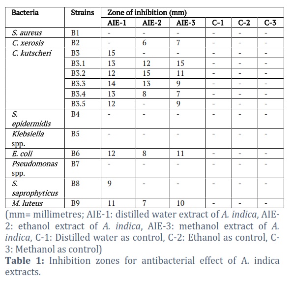
Among A. indica extracts, none showed any activity against S. aureus, for C. xerosis AIE-2 and AIE-3 showed inhibition zones of 6mm and 7mm respectively and no zone with AIE-1. With C. kutscheri AIE-1 gave zones of 15mm, 13mm, 12mm, 14mm, 13mm and 12mm against B3, B3.1, B3.2 and B3.3, B3.4 and B3.5 respectively. For strain B3 no zone with AIE-2 and AIE-3 was obtained while for strain B3.1, B3.2 and B3.3, B3.4 and B3.5 zones of 12mm, 15mm, 13mm, 8mm, 0mm with AIE-2 and zones of 15mm, 11mm, 9mm, 7mm and 9mm with AIE-3 were obtained, respectively. No extract gave any zones against S. epidermidis, Klebsiella spp. and Pseudomonas spp. in case of A. indica. Zones of 122mm, 8mm and 11mm were obtained against E. coli and for M. luteus 11mm, 7mm, and 10mm zones were observed on the plates with AIE-1, AIE-2, and AIE-3 extracts respectively. Only a single zone of 9mm was obtained against S. saprophyticus with AIE-1. Controls (C-1, C-2, C-3) used were respective solvents and they produced no zones of inhibition. Table 1 holds the results for A. indica extracts against the selected bacterial strains.
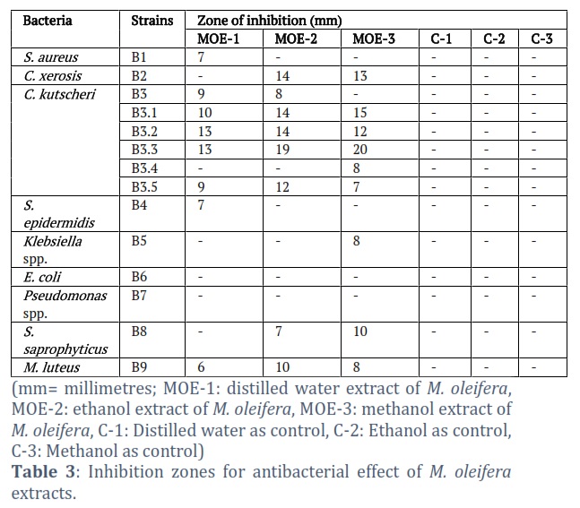
No zone at all was obtained with M. oleifera extracts against E. coli and Pseudomonas spp. Only MOE-1 gave a zone of 7mm against both S. aureus and S. epidermidis while no zone appeared with either MOE-2 or MOE-3. Zone of 13mm and 14mm were produced against C. xerosis with MOE-2 and MOE-3 respectively. Zones of 9mm, 10mm, 13mm and 13mm with MOE-1, 8mm, 14mm, 14mm,19mm, 9mm and 12mm with MOE-2 were obtained against B3, B3.1, B3.2, B3.3, B3.4 and B3.5 respectively against C. kutscheri. MOE-3 gave no inhibition zone for B3 but 15mm against B3.1, 12mm against B3.2, 20mm zones against B3.3, 8mm zone against B3.4 and 7mm zone against B3.5. A single zone of 8mm with MOE-3 was obtained against Klebsiella spp. and none with MOE-1 and MOE-2. Zones of 7mm and 10mm were obtained with MOE-2 and MOE-3 respectively against S. saprophyticus while MOE-1 produced no zone against it. Against M. luteus zones of 6mm, 10mm and 8mm were obtained with MOE-1, MOE-2 and MOE-3 respectively. Controls (C-1, C-2, C-3) used were respective solvents and they produced no zones of inhibition. Table 3 contains information on M. oleifera extracts.
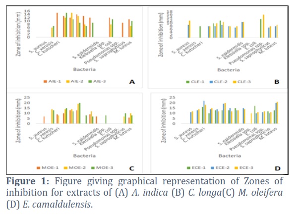
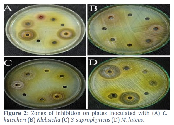
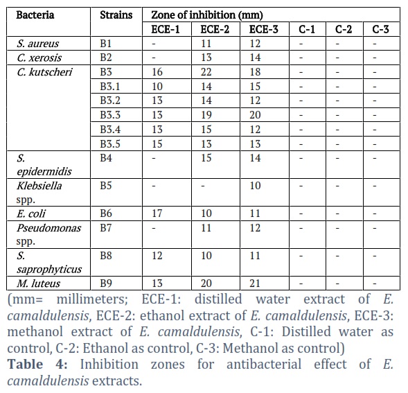
Among the E. camaldulensis extracts, excluding some of the extracts almost all of them gave positive results in the form of inhibition zones. Against S. aureus ECE-2 and ECE-3 produced zones of 11mm and 12mm respectively while ECE-1 gave no zone. 13mm and 14mm clear zones appeared with ECE-2 and ECE-3 respectively against C. xerosis while no zone was observed by ECE-1. In case of C. kutscheri 16mm, 10mm, 13mm, 13mm, 13mm,15mm zones with ECE-1, 22mm, 14mm, 14mm, 19mm, 15mm, 13mm zones with ECE-2 and 18mm, 15mm, 12mm, 20mm, 12mm, 13mm zones with ECE-3 were formed against B3, B3.1, B3.2, B3.3, B3.4 B3.5 strain respectively. When tested against S. epidermidis ECE-2 and ECE-3 generated two zones of 15mm and 14 mm respectively while ECE-1 made none. Only ECE-3 gave a clear 10mm zone against Klebsiella spp. while growth remained unbothered by ECE-1 and ECE-2. Clear zones of 17mm, 10mm and 11mm were formed with ECE-1 ECE-2 and ECE-3 on E. coli plates. 11mm and 12mm zone with ECE-2 and ECE-3 against Pseudomonas spp., 12mm, 10mm, 11mm clear zones with ECE-1, ECE-2 and ECE-3 against S. saprophyticus and 13mm, 20mm, 21mm clear zones with ECE-1, ECE-2, ECE-4 against M. luteus were obtained. Controls (C-1, C-2, C-3) used were respective solvents and they produced no zones of inhibition. Table 4 comprises of the test results for testing antibacterial potential of E. camaldulensis with figure 1 (A, B, C, D) giving a graphical representation of inhibition zones for all four plants. Figure 2 (A, B, C and D) shows some inhibition zones obtained on various bacterial plates.
Figures & Tables
As increasing degree of bacterial resistance and emergence of pan- and multi-drug resistant bacteria has become a global issue, the world is turning to exploring the plant kingdom in need of new antimicrobial agents and treatment strategies [4,31,32], Plants owe these medicinal properties to the presence of many bioactive substances which render plants with various biological potentials namely reduction, anti-microbial, anti-oxidant, anti-coagulant, anti-tumor, anti-diabetic, anti-inflammatory etc [1,16] .
In current study four plants namely Azadirachta indica, Curcuma longa, Moringa oleifera and Eucalyptus camaldulensis were selected and their extracts were prepared to study their antimicrobial effects on human skin flora. Leaves of A. indica, M. oleifera and E, camaldulensis while rhizome of C. longa was collected for this study, and after proper cleaning these were set to dry in shade under ambient conditions. Dried material was powdered and extracts were prepared using distilled water, 50% ethanol and 50% methanol as solvents. Presence of phytoconstituents namely saponins, proteins, tannins, alkaloids, flavonoids, phenols, steroids/terpenoids, and carbohydrates was determined qualitatively in each extract and their antibacterial potential was then evaluated by agar well diffusion assay.
In current study, extract of A. indica prepared with ethanol as solvent contains highest amount of phytoconstituents followed by distilled water and methanol solvents as they lack tannins saponins respectively as mentioned below. AIE-1 was found to contain all the above-mentioned phytochemicals which was basically in accordance with the findings of [33] with the exception of tannins as they were not found here in current study. All of these phytochemicals were present in AIE-2 in our study while another study reported the presence of some with absence of saponins, protein and alkaloids in extracts with same solvent [34] . AIE-3 in this study was only lacking in the presence of saponins and this finding was majorly compatible with the findings of [33] where the only exception was presence of saponins.
C. longa extract for which distilled water served as a solvent contained five out of eight tested phytochemicals while those prepared with ethanol and methanol contained four of them only as is explained further. CLE-1 was found to carry saponins, alkaloids flavonoids, steroids/terpenoids and carbohydrates with absence of proteins, tannins and phenols while [35] reported the same extracts to carry phenols and absence of saponins and terpenoids, but another study reported the same extract with absence of alkaloids, saponins and presence of tannins [36] yet another study reported the absence of tannins and phenols and presence of saponins, alkaloid,, flavonoids, terpenoids and carbohydrates as is the case in our study [37] . CLE-2 showed only the presence of alkaloids, flavonoids, steroids/ terpenoids and carbohydrates with absence of all others in current study while another study reported the presence of alkaloids and terpenoids along with phenols, tannins and absence of flavonoids and carbohydrates [1] . CLE-3 in current study also showed the presence of alkaloids, flavonoids, steroids/terpenoids and carbohydrates. This presence was pretty much in accordance with the findings of another study but absence of phytochemicals reported in our study was contradictory due to their presence in study of [37] Yet another study showed almost similar results with exception of the results for flavonoids, phenols and saponins in the same extracts [1] .
For M. oleifera, extracts prepared in ethanol and methanol solvents indicated the presence of all phytoconstituents but extract prepared in distilled water was lacking in one of these. MOE-1 showed the presence of all aforementioned phytochemicals with exception of steroids/terpenoids which coincides with the finding of [14] . MOE-2 contained all the phytochemicals for which the tests were performed in our study while another study reported ethanol extract that was lacking flavonoids and steroids [38,39] . MOE-3 was also found to contain all the tested phytochemicals in current study and this was compatible with the findings in a study [40] . Aqueous and ethanolic extracts of E. camaldulensis i.e. ECE-1and ECE-2 showed the presence of all phytochemicals that were tested in our study, this result was fairly compatible with the findings of a study but was contradicting regarding the presence of alkaloids that were reported absent by them [41] . Another study reported the aqueous extracts to carry most of the under-study phytochemicals with a few namely proteins, steroids and alkaloids as undetected [42] .
Methanolic extracts of Eucalyptus camaldulensis ECE-3 were found to carry all phytochemicals but saponins in our study and these results were highly in accordance with the findings of a study conducted by [43] that also reported these phytochemicals to be present in E. camaldulensis extracts. Our results also align with the findings of [44] with proteins being the undetected part. Among A. indica extracts, no zone of inhibition against S. aureus, S. epidermidis, Klebsiella spp. and Pseudomonas spp. at all was present irrespective of the solvent used while a study reported the inhibition of growth of S. aureus Klebsiella spp. and Pseudomonas spp. by methanol extract [45] . Another study conducted by [46] concluded that aqueous and ethanol extracts of neem formed zones of inhibition against S. aureus, S. epidermidis and Pseudomonas spp. reported aqueous neem extracts to be effective against Klebsiella spp. and Pseudomonas spp. Only AIE-1 showed activity against S. saprophyticus and none of the other two extracts. Ethanolic extract i.e., AIE-2 showed zones of various diameters against C. xerosis and strains of C. kutscheri.
All the extracts irrespective of the solvent showed zones of inhibition against E. coli, M. luteus which tells that E.coli and M. luteus are the most sensitive strains under study that respond to A. indica extracts irrespective of the solvent and this finding is compatible with the findings of various other studies where E.coli growth was inhibited by A. indica aqueous extracts [46] ethanol extract [36] and methanol extracts [45] yet another study reported that irrespective of the solvent used for A. indica extract preparation, no zone of inhibition against E.coli appeared and this was contrary to our findings [47] .
Our findings were contradicting with the findings of another study where aqueous and ethanol extracts inhibited the growth of S. aureus at different concentrations of extracts while no zones against S. aureus appeared in our study [48] . No results regarding S. saprophyticus, C. xerosis and C. kutscheri were found in the searched literature and this might be because other bacteria are common findings as compared to these three.
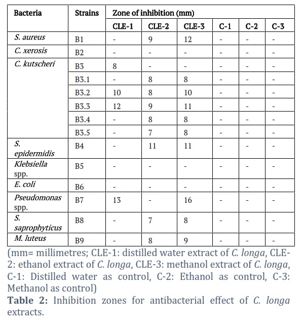
Among C. longa extracts, no zone of inhibition against C. xerosis, Klebsiella spp. and E. coli was formed irrespective of the solvent used in our study which contradicted with the findings of the study where aqueous and ethanolic extracts inhibited the activity of E. coli and Klebsiella spp. [35] . In a study methanolic extracts of turmeric were found to hinder the growth of E. coli [49] and yet another study [34] reported that methanolic extracts of turmeric have growth inhibitory potential against E. coli and Klebsiella spp., but our findings were compatible with the results of [16] where aqueous extract of C. longa showed no zone of inhibition against Klebsiella spp. and E. coli and ethanolic extract also showed no activity against Klebsiella spp. CLE-1 in current study showed zones of inhibition with varying diameters against three out of six C. kutscheri strains and Pseudomonas spp. which contradicts with result of same extract showing no activity against Pseudomonas spp. [35] and is compatible with the finding of [1] where growth of Pseudomonas spp. was hindered by the same extract. It made no zone against S. aureus, S. epidermidis, S. saprophyticus and M. luteus while it showed zone of inhibition against S. aureus [35] and against S. aureus and S. epidermidis in the study of [1] . CLE-2 in our study showed zones of inhibition with almost similar diameters against S. aureus, five out of six C. kutscheri strains, S. epidermidis, S. saprophyticus and M. luteus and no such zone against Pseudomonas spp. It falls in accordance with the results of many studies which concluded in same extract showing activity against S. aureus [49-51] while contradict with some studies where activity is exhibited against Pseudomonas spp. [1,21] .
In this study CLE-3 was also presented with inhibitory effects by showing inhibition zones against S. aureus, five out of six C. kutscheri strains, S. epidermidis, Pseudomonas spp. S. saprophyticus and M. luteus which is in accordance with the findings of [21] . Out of all the M. oleifera extracts, neither showed any zone of inhibition against E. coli and Pseudomonas species which contradicts with the results stated in a study [22] where activity against E. coli and Pseudomonas spp. were exhibited by ethanol extracts of same plant along-with another study where aqueous as well as methanolic extracts showed activity against E. coli [52] . But the findings are pretty much in accordance with the results of a study where M. oleifera aqueous extracts exhibited no zones of inhibition against E. coli, Pseudomonas spp. and Klebsiella spp. and S. aureus [53] . MOE-1 showed a single zone against S. aureus which is similar to the findings of a study where aqueous extract also showed antibacterial potential against S. aureus [51] . MOE-2 gave no clear zone against either S. aureus or Klebsiella spp. while in another study it did produces clear zones against Klebsiella as reported by [12] . A single zone against Klebsiella spp. was exhibited by MOE-3 and none against S. aureus while zones against S.aureus were exhibited by methanolic M. oleifera extracts in a study [52] . Zones of inhibition against C. kutscheri, S. epidermidis, M. luteus by MOE-1, against C. xerosis, C, kutscheri, S. saprophyticus, M. luteus by MOE-2 and against C. xerosis, C. kutscheri, S. saprophyticus and M. luteus by MOE-3 were also formed in recent study but were not discussed because no data about the effect of these extracts on aforementioned bacteria was found on repeated search.
In current study, at least some extent of antibacterial potential by at least one of the extracts of E. camaldulensis was exhibited against each selected bacterial strains. ECE-1 did not show any zone of inhibition against S. aureus, Klebsiella spp. and Pseudomonas spp. which was contrary to the results stated in other studies where Klebsiella spp. and Pseudomonas spp. growth was inhibited by utilizing same extracts in these studies [1,37] . [21] reported E. camaldulensis aqueous extract to be active against Klebsiella and S. aureus, while a different study concluded that no zone of inhibition was formed by aqueous extract of this plant against S. aureus [54] . ECE-1 exhibited inhibitory zones against E. coli. These outcomes were compatible with the result of other studies about the activity against E. coli [18] . ECE-2 showed antibacterial potential through zones of varying diameters against S. aureus, E. coli, and Pseudomonas spp. and these results aligned with the findings in some studies. While occurrence of no zone against Klebsiella spp. in our study was contradictory to the findings of some other studies [15,33] . ECE-3 formed zones of inhibition against S. aureus, Klebsiella spp., E. coli and Pseodomonas spp. which was contradictory to the findings of [11] because they showed methanolic extract to make no zone against S. aureus and compatible in showing a zone of inhibition against Pseudomonas spp. respectively. It was also compatible with the findings of another study where zone of inhibition against Klebsiella was observed [55] . Zones against C. kutscheri, S. saprophyticus and M. luteus were also formed irrespective of the solvent used while ECE-2 and ECE-3 gave zone of inhibition against C. xerosis also. Data on the effect of extracts against these bacteria was surprisingly not found in any of the searched paper and literature for all the plants which makes the inclusion of these bacteria, a novelty for our paper.
Statistical analysis performed for extracts of each plant showed p value for A. indica as 0.36 (P > 0.05), for C. longa as 0.09 (P > 0.05), for M. oleifera as 0.67 (P > 0.05) and for E. camaldulensis as 0.02 (P < 0.05), which indicates significant antibacterial activity of E. camaldulensis depending on the size of zones of inhibition produced by it. Most of the human skin flora included in this study namely Staphylococcus aureus, Staphylococcus epidermidis, Corynebacterium xerosis, Corynebacterium kutscheri, Klebsiella spp., Escherichia coli, Pseudomonas spp., Staphylococcus saprophyticus, and Micrococcus luteus that can also act as opportunistic pathogens was susceptible to different type of extracts prepared from A. indica, C. longa, M. oleifera and E. camaldulensis by varying degrees. Thus our study proves the use of various extracts prepared from these plants for antibacterial purposes after careful evaluation of concentration, type and presence or absence of specific phytochemicals required for the action. This study also suggests the use of these extracts for evaluation of further health potentials e.g., anti- diabetic, anti-oncogenic and anti-oxidant etc. As the results are based on qualitative analysis, the study further suggests investigations related to different concentrations e.g., by dilutions of these extracts that might be helpful in harnessing the maximum effects based on the most effective concentration. The study also suggests investigations about the Molecular mechanism of action of these extracts and changes in the target microorganisms.
Conflict of Interest
The authors declare that there is no conflict of interest.
study conception and design: Rabeea Anwar, Humaira Yasmeen; data collection: Rabeea Anwar; contributed data or analysis tools: Zeshan Ali; performed the analysis and interpretation of results: Rabeea Anwar, Saigha Marriam; draft manuscript preparation: Rabeea Anwar; Revised it critically for important intellectual content: Zeshan Ali, Muhammad Nauman Sharif; Supervised the whole study and experimental work and approved for further proceeding: Zeeshan Ali, Humaira Yasmeen, Iqra Arooj; All authors reviewed the results and approved the final version of the manuscript.
![]()
References
- Khan S, Ur-Rehman T, Mirza B, Ul-Haq I, Zia M. Antioxidant, antimicrobial, cytotoxic and protein kinase inhibition activities of fifteen traditional medicinal plants from Pakistan. Pharmaceutical Chemistry Journal, (2017); 51391-398.
- Ahmed N, Karobari MI, Yousaf A, Mohamed RN, Arshad S, et al. The Antimicrobial Efficacy Against Selective Oral Microbes, Antioxidant Activity and Preliminary Phytochemical Screening of Zingiber officinale. Infection and Drug Resistance, (2022); 15: 2773-2785.
- Yadav R, Agarwala M. Phytochemical analysis of some medicinal plants. Journal of phytology, (2011); 3(12): 10-14.
- Ramzan M, Karobari MI, Heboyan A, Mohamed RN, Mustafa M, et al. Synthesis of silver nanoparticles from extracts of wild ginger (Zingiber zerumbet) with antibacterial activity against selective multidrug resistant oral bacteria. Molecules, (2022); 27(6): 2007.
- Javed F, Ali Z, Ali S, Ahmed N, Alam MK, et al. BARLEY BRAN, A NOVEL AGRICULTURAL WASTE FOR THE IMPROVED PRODUCTION OF AN EXTRACELLULAR LACCASE FROM A SOIL-INHABITED Penicillium spp. Journal of microbiology, biotechnology and food sciences, (2022); e3631-e3631.
- Tariq F, Ahmed N, Afzal M, Khan MAU, Zeshan B. Synthesis, Characterization and antimicrobial activity of Bacillus subtilis-derived silver nanoparticles against multidrug-resistant bacteria. Jundishapur Journal of Microbiology, (2020); 13(5).
- Saleem S, Muhammad G, Hussain MA, Bukhari SNA. A comprehensive review of phytochemical profile, bioactives for pharmaceuticals, and pharmacological attributes of Azadirachta indica. Phytotherapy research, (2018); 32(7): 1241-1272.
- Ali Z, Jatoi MA, Al-Wraikat M, Ahmed N, Li J. Time to enhance immunity via functional foods and supplements: Hope for SARS-CoV-2 outbreak. Altern Ther Health Med, (2021); 2730-44.
- Shinwari ZK, Qaiser M. Efforts on conservation and sustainable use of medicinal plants of Pakistan. Pak J Bot, (2011); 43(1): 5-10.
- Hasan Z, Zeshan B, Hassan A, Daud NHA, Sadaf A, et al. Preparation and characterization of edible whey protein nanofibrils and efficacy studies on the quality and shelf‐life of chilled food products. Journal of Food Safety, (2022); e13034.
- Ahmad S, Maqbool A, Srivastava A, Gogol S. Biological detail and therapeutic effect of azadirachta indica (neem tree) products-a review. Evidence Based Med Healthcare, (2019); 6(22): 1607-1612.
- Saleem A, Ahotupa M, Pihlaja K. Total phenolics concentration and antioxidant potential of extracts of medicinal plants of Pakistan. Zeitschrift für Naturforschung C, (2001); 56(11-12): 973-978.
- Rajkumari S, Sanatombi K. Nutritional value, phytochemical composition, and biological activities of edible Curcuma species: A review. International journal of food properties, (2017); 20(sup3): S2668-S2687.
- Dhakad AK, Ikram M, Sharma S, Khan S, Pandey VV, et al. Biological, nutritional, and therapeutic significance of Moringa oleifera Lam. Phytotherapy Research, (2019); 33(11): 2870-2903.
- Gandji K, Chadare F, Idohou R, Salako V, Assogbadjo A, et al. Status and utilisation of Moringa oleifera Lam: A review. African Crop Science Journal, (2018); 26(1): 137-156.
- Sabo VA, Knezevic P. Antimicrobial activity of Eucalyptus camaldulensis Dehn. plant extracts and essential oils: A review. Industrial crops and products, (2019); 132413-429.
- Ghareeb MA, Habib MR, Mossalem HS, Abdel-Aziz MS. Phytochemical analysis of Eucalyptus camaldulensis leaves extracts and testing its antimicrobial and schistosomicidal activities. Bulletin of the National Research Centre, (2018); 421-9.
- Abubakar AR, Haque M. Preparation of medicinal plants: Basic extraction and fractionation procedures for experimental purposes. Journal of pharmacy & bioallied sciences, (2020); 12(1): 1.
- Roghini R, Vijayalakshmi K. Phytochemical screening, quantitative analysis of flavonoids and minerals in ethanolic extract of Citrus paradisi. International Journal of Pharmaceutical Sciences and Research, (2018); 9(11): 4859-4864.
- Sasidharan S, Chen Y, Saravanan D, Sundram K, Latha LY. Extraction, isolation and characterization of bioactive compounds from plants’ extracts. African journal of traditional, complementary and alternative medicines, (2011); 8(1): 1-10.
- Akbar A, Ali I, Samiullah NU, Khan SA, Rehman Z, et al. Functional, antioxidant, antimicrobial potential and food safety applications of curcuma longa and cuminum cyminum. Pakistan Journal of Botany, (2019); 51(3): 1129-1135.
- Parveen S, Saqib S, Ahmed A, Shahzad A, Ahmed N. Prevalence of MRSA colonization among healthcare-workers and effectiveness of decolonization regimen in ICU of a Tertiary care Hospital, Lahore, Pakistan. Advancements in Life Sciences, (2020); 8(1): 38-41.
- Sohail M, Muzzammil M, Ahmad M, Rehman S, Garout M, et al. Molecular Characterization of Community-and Hospital-Acquired Methicillin-Resistant Staphylococcus aureus Isolates during COVID-19 Pandemic. Antibiotics, (2023); 12(1): 157.
- Rizvi A, Saeed MU, Nadeem A, Yaqoob A, Rabaan AA, et al. Evaluation of Bi-Lateral Co-Infections and Antibiotic Resistance Rates among COVID-19 Patients in Lahore, Pakistan. Medicina, (2022); 58(7): 904.
- Ahmed N, Khalid H, Mushtaq M, Basha S, Rabaan AA, et al. The Molecular Characterization of Virulence Determinants and Antibiotic Resistance Patterns in Human Bacterial Uropathogens. Antibiotics, (2022); 11(4): 516.
- Ahmed N, Khan M, Saleem W, Karobari M, Mohamed R, et al. Evaluation of Bi-Lateral Co-Infections and Antibiotic Resistance Rates among COVID-19 Patients. Antibiotics (2022); (11): 276.
- Ahmed N, Zeshan B, Naveed M, Afzal M, Mohamed M. Antibiotic resistance profile in relation to virulence genes fimH, hlyA and usp of uropathogenic E. coli isolates in Lahore, Pakistan. Trop Biomed, (2019); 36559-568.
- Ahmed N, Tahir K, Aslam S, Cheema SM, Rabaan AA, et al. Heavy Metal (Arsenic) Induced Antibiotic Resistance among Extended-Spectrum β-Lactamase (ESBL) Producing Bacteria of Nosocomial Origin. Pharmaceuticals, (2022); 15(11): 1426.
- Ahmed N, Ali Z, Riaz M, Zeshan B, Wattoo JI, et al. Evaluation of antibiotic resistance and virulence genes among clinical isolates of Pseudomonas aeruginosa from cancer patients. Asian Pacific journal of cancer prevention: APJCP, (2020); 21(5): 1333.
- Zeshan B, Karobari MI, Afzal N, Siddiq A, Basha S, et al. The usage of antibiotics by COVID-19 patients with comorbidities: the risk of increased antimicrobial resistance. Antibiotics, (2021); 11(1): 35.
- Ahmad A, Baig AA, Hussain M, Saeed MU, Bilal M, et al. Narrative on Hydrogen Therapy and its Clinical Applications: Safety and Efficacy. Current Pharmaceutical Design, (2022); 28(31): 2519-2537.
- Al-Hatamleh MA, Alshaer W, Ma’mon MH, Lambuk L, Ahmed N, et al. Applications of Alginate-Based Nanomaterials in Enhancing the Therapeutic Effects of Bee Products. Frontiers in Molecular Biosciences, (2022); 9.
- Hanif H, Elikaei A, Vazini H, Mohammadi A. Anticancer and Antibacterial Effect of Eucalyptus Camaldulensis, in Vitro. Medical Laboratory Journal, (2021); 15(1): 26-32.
- Sakha H, Hora R, Shrestha S, Acharya S, Dhakal D, et al. Antimicrobial activity of ethanolic extract of medicinal plants against human pathogenic bacteria. Tribhuvan University Journal of Microbiology, (2018); 51-6.
- Jassal PS, Thambyrajah JC. Antibacterial and phytochemical analysis of condiments. Drug Invention Today, (2018); 10(5): 844-847.
- Hosea ZY, Kator L, Rhoda EH. Phytochemical properties and antimicrobial activities of aqueous extract of Curcuma longa (Turmeric) rhizome extract. Asian Journal of Research in Crop Science, (2018); 2(1): 1-8.
- Pandey J, Mishra S, Jaiswal K. In vitro evaluation of the anthelmintic activity of rhizome extracts of Curcuma Longa (Linn.). IN VITRO, (2018); 11(12).
- Oluwajobi I, Kabiru YA, Jigam AA. Antibacterial and Antifungal activities of aqueous leaves extract of some medicinal plants. GSC Biological and Pharmaceutical Sciences, (2019); 9(1): 062-069.
- Al-Mhanna SB, Ghazali WSW, Mohamed M, Rabaan AA, Santali EY, et al. Effectiveness of physical activity on immunity markers and quality of life in cancer patient: a systematic review. PeerJ, (2022); 10e13664.
- Negassa Z, Furgasa W, Mekonnen N. Phytochemical screening and in vitro anthelminthic activity of selected ethnoveterinary medicinal plants in Haramaya District, Eastern Hararghe zone, Ethiopia.
- Ishag OAO, Erwa IY, Diriye MA, Lawane AAM, Ahmed HM, et al. antimicrobial potential and phytochemical screening of Eucalyptus camaldulensis and Eucalyptus microtheca leaves extracts. South Asian Research Journal of Natural Products, (2018); 1(3): 1-6.
- Anigboro AA, Avwioroko OJ, Cholu CO. Phytochemical constituents, antimalarial efficacy, and protective effect of Eucalyptus camaldulensis aqueous leaf extract in plasmodium berghei-infected mice. Preventive nutrition and food science, (2020); 25(1): 58.
- Haruna H, Gabi B, Umar H. Phytochemical Analysis and Anti-Malarial Potential of Eucalyptus camaldulensis (Turare) Leaf Fractions.
- Kaur S, Gupta S, Gautam PB. Phytochemical analysis of Eucalyptus leaves extract. Journal of Pharmacognosy and Phytochemistry, (2019); 8(1): 2442-2446.
- Nigussie D, Davey G, Legesse BA, Fekadu A, Makonnen E. Antibacterial activity of methanol extracts of the leaves of three medicinal plants against selected bacteria isolated from wounds of lymphoedema patients. BMC Complementary Medicine and Therapies, (2021); 211-10.
- Ali MJ, Obaid RF, Obaid RF. Antibacterial activity for acne treatment through medicinal plants extracts: novel alternative therapies for acne. Journal of Pure and Applied Microbiology, (2019); 13(2): 1245-1250.
- Saxena A, Mukhopadhyay A, Nandi S. Antibacterial activity of selected plants extract against pathogenic bacteria and detection of phytochemicals. Journal of Environmental Biology, (2020); 41(6): 1486-1492.
- Yelmate A, Thonte S. Antibacterial activity of some Indian medicinal plants against methicillin resistant Staphylococcus aureus (MRSA). Journal of Pharmacognosy and Phytochemistry, (2019); 8(5): 376-380.
- Irshad S, Muazzam A, Shahid Z, Dalrymple MB. Curcuma longa (Turmeric): An auspicious spice for antibacterial, phytochemical and antioxidant activities. Pak J Pharm Sci, (2018); 31(6): 2689-2696.
- Hossaini F, Das NC, Hossaini F, Acharjee M, Munshi SK. Antimicrobial traits of different medicinal plants locally available in Bangladesh. Biomedical and Biotechnology Research Journal (BBRJ), (2021); 5(1): 1.
- Husein HA, Alhasan DA, Albadry MA. In Vitro Antimicrobial Activity and GC-MS Analysis of Crude Aqueous Methanolic Extract Produced from Leaves of Eucalyptus species. Med J, (2019); 172019.
- Garga M, Garasin U, Abdullahi M, Muhammed B, Yakubu A, et al. ANTIBACTERIAL ACTIVITY AND PHYTOCHEMICAL SCREENING OF MANGIFERA INDICA ETHANOL AND AQUEOUS LEAVES EXTRACT AGAINST PSEUDOMONAS AERUGINOSA AND STAPHYLOCOCCUS AUREUS.
- Seddiek AS, Hamad GM, Zeitoun A, Zeitoun M, Ali S. Antimicrobial and antioxidant activity of some plant extracts against different food spoilage and pathogenic microbes. Eur J Nutr Food Saf, (2020); 121-12.
- Al-Hadidy YI, Yaseen SS, Saleh GM. The Inhibitory Effect of some Plant Extracts on some Pathogenic Bacteria. Tikrit Journal of Pure Science, (2019); 24(1): 62-69.
- Chiamaka OS, Ndarubu TA, Mahmood MF, Adenike AR, Alfa S, et al. In Vitro Antioxidants, Antimicrobials and Biochemical Response of Methanol Leaf Extract of Eucalyptus camaldulensis following Sub-Acute Administration to Rats. Saudi Journal of Biomedical Research (2019); 4(11): 405-411.
This work is licensed under a Creative Commons Attribution-Non Commercial 4.0 International License. To read the copy of this license please visit: https://creativecommons.org/licenses/by-nc/4.0
![]()


