

Full Length Research Article
Bioactivity and Metabolomics Profiling of Endophytic Actinobacteria Isolated from Roots of the Medicinal Plants Dominant in South Asian Region
Ashba Hassan1,2,3, Larissa V. Ponomareva2,3, Jon S. Thorson2,3, Khaled A. Shaaban2,3, Imran Sajid1,2,3,*
Adv. life sci., vol. 10, no. 2, pp. 200-209, June 2023
*– Corresponding Author: Imran Sajid (imran.mmg@pu.edu.pk)
Authors' Affiliations
2.Center for Pharmaceutical Research and Innovation (CPRI) College of Pharmacy, University of Kentucky, Lexington, Kentucky – USA
3. Department of Pharmaceutical Sciences, College of Pharmacy, University of Kentucky, Lexington, Kentucky – USA
[Date Received: 27/02/2022; Date Revised: 21/03/2023; Date Published Online: 30/06/2023; Date Updated:11/09/2025]
Abstract![]()
Introduction
Methods
Results
Discussion
References
Abstract
Background: Plant-derived endophytic actinobacteria are the center of attention due to their capacity to produce diverse antimicrobial and anticancer compounds and their metabolites influence plant growth.
Methods: In this study, 40 endophytic actinobacteria strains were isolated from the roots of eight medicinal plants used as folk medicine in South Asian region. The isolates were characterized morphologically, biochemically and physiologically and the genus level identification of the selected strains was done by 16SrRNA gene sequencing. In small scale cultivation (50ml broth), the isolates were grown in A-medium to prepare the crude extracts. These crude extracts were subsequently evaluated for their antimicrobial, anticancer and antioxidant activity and the metabolomics profile of each of the extract was determined by TLC and HPLC-UV/MS.
Results: The taxonomic studies showed that the isolates belong to the group actinobacteria based on their morphological and physiological characteristics and the 16SrRNA gene sequencing of the selected strains identified the genera including Streptomyces, Micromonospora and Nocardia. Cumulatively,53% of extracts exhibited anti-Gram-(+) activity,47% exhibited anti-Gram-(-) activity,32% exhibited antifungal activity and 30% were cytotoxic to PC3 and A549 cancer cell lines and most of the extracts have shown antioxidant activity greater than 50%. The metabolomics analysis predicted the presence of an array of low molecular weight metabolites and indicated the promising isolates in collection for further studies for novel bioactive metabolite isolation and structure elucidation.
Conclusion: Overall the study provides an overview of the endophytic actinobacteria residing in the roots of the selected medicinal plants prevalent in south Asian region and their potential to produce the medicinally and biotechnologically useful compounds.
Keywords: Endophytic Actinobacteria; Metabolomic Profiling; Natural Products; Antimicrobial Compounds; Anticancer Compounds
Introduction![]()
Plant derived bioactive compounds and their metabolic byproducts, or the secondary metabolites produced by the residential endophytic microbes (microbes living inside the plant tissues) have been the rich source of bioactive metabolites. The root exudates secreted by the plants attract microbes from their surroundings [1]. The endophytic microbial flora benefit plants with respect to ecological and evolutionary perspective [2]. Their mutualistic association influences the fitness of the plants by various mechanisms, including the induction of systemic resistance [3], and improves the plant health [4]. Secondary metabolites produced by endophytic bacteria also contribute to the heterogeneity of the phytochemical profile of the plants [5]. The soil harbors enormous diversity of various microbial groups, among them actinobacteria are the most abundant soil dwelling bacteria and are present in almost all types of soils. The association of the actinobacteria with roots is common as compared to other plant parts due to the close proximity of the roots with soil microbes (rhizospheric soil population) [6] and due to the chemical cues secreted by plants [7].
The increasing focus on the screening of actinobacteria from diverse and unusual environments has made them attractive targets for medicinally useful compounds. For example, endophytic actinobacteria from mangrove plants have been reported to produce 84 new compounds including salinosporamides, xiamycin and novel indolocarbazoles [8].Xu et al. [9] hypothesized endophytic actinobacteria to provide a greater percentage of novel metabolites and revealed 73 novel compounds from such sources. Among the endophytic actinobacteria, the genus Streptomyces accounts for the abundant genus in plant tissues followed by various other genera [10]. The endophytic Glycomyces and Streptomyces have proved to be the producer of bioactive compounds against penicillin resistant S. aureus [11]. The plant growth promoting compounds produced by the endophytic actinobacteria can enhance the growth of host plant [12]. In 2002, Igarashi et al. [13] reported 398 endophytic actinomycetes having antagonistic activity against phytopathogens. The 2 new novobiocin antibiotics were obtained from the endophytic Streptomyces isolated from the Aucuba japonica [14]. Two novel butyrolactone antibiotics (cedarmycin A and B) were derived from the endophytic Streptomyces isolated from Cryptomeria japonica [15]. Alnumycin, a new naphthoquinone antibiotic was reported from the endophytic Streptomyces isolated from the Alnus glutinosa [16]. Endophytic actinobacteria not only produce bioactive secondary metabolites but also show positive effect on plant growth.
The isolates from chickpeas have shown not only positive effect on its growth but also protected the plant from pathogen and produced industrially important enzymes (chitinase, cellulase and protease) [17]. Similarly, the endophytic strain Jishengella endophytica have been reported to produce the 1-hydroxy-Beta-carboline against influenza virus type A subtype H1N1 [18].
In this study, the root associated endophytic actinobacteria were isolated from 8 selected medicinal plants. A comprehensive identification strategy including morphological, physiological, biochemical and genomic characterization of the isolates was adopted. The crude extracts of the isolated strains exhibited promising antimicrobial activity against the bacterial test strains, also high cytotoxicity/anticancer activity against the human cancerous cell lines and significant antioxidant activity was observed in in-vitro assays. Similarly, the extracts exhibited impressive metabolomic profiles and presence of low molecular weight compounds by TLC HPLC-UV/MS analysis. Based on the bioactivity and chemical profiling, 7 isolates were prioritized including PU-Aml3, PU-LG3, PU-Aki2, PU-Aki3, PU-Aki4, PU-Kach5 and PU-Kach15 for future scale-up fermentation, compounds purification and structure elucidation.
Methods![]()
Sample Collection
The root samples were collected in sterile plastic bags from various locations in the botanical garden and adjoining areas at the campus of University of the Punjab, Lahore, Pakistan and were transferred to the laboratory for further processing. The sampled plants included Cassia fistula, Phyllanthus emblica, Bauhinia variegate, Cassia occidentalis, Cymbopogon, Ocimum tenuiflorum, Moringa oleifera and Tamarindus indica.
Surface Sterilization and Selective Isolation of the Endophytic Actinobacteria
The root samples of plants were surface sterilized using chemical sterilant of different concentrations with a slight modification to the original surface sterilization protocol by Cao et al. [19]. The collected roots were thoroughly washed with tap water and then sequentially sterilized by i) 70% ethanol for 5 min and rinsed with sterile water ii) 0.1% sodium hypochlorite solution for 10min and rinsed with sterile water and iii) 70% ethanol for 5 min, iv) rinsed (x4) with sterile water. The surface sterilized roots were fragmented into small pieces of 2-3cm in size using sterilized scalpel and the fragments were placed slightly embedded on actinomycetes isolation agar (Difco), starch-casein-KNO3 agar and raffinose-histidine agar. The efficacy of the surface sterilization procedure was evaluated by negative control media plate which was inoculated with 60 µl of last wash of water used in sterilization protocol and incubated at 28°C for 3-4 weeks. The colonies of actinobacteria that appeared on the isolation agar were further sub-cultured on A-media. The preliminary phenotypic characterization was done by following the guidelines as mentioned in Bergey’s manual of systematic bacteriology Volume II for initial identification of the isolates [20].
Taxonomic Identification of the Endophytic Actinobacteria
The endophytic actinobacteria retrieved from the roots embedded on the agar were distinguished from the fungus by visualizing the plates under stereomicroscope. The phenotypic identification of isolates included macroscopic characterization (color of substrate and aerial mycelium, pigment production and texture of the colonies) and microscopic examination included gram staining, while the biochemical characterization was done by determining, utilization of various carbon and nitrogen sources, melanin production, esculin hydrolysis [21,22]. The production of amylase, pectinase and cellulase enzymes was determined following the protocols of Minotto et al. [23]. The optimal conditions for the growth of endophytic actinobacteria were determined under a range of temperature (28°C, 37°C and 45°C) and pH conditions [7-9, 11]. The indole production was analyzed by the protocol of Yandigeri et al. [24], and phosphate solubilization activity was determined on Pikovskaya medium [25].The 16S rRNA gene sequences from forward and reverse primers were assembled and the resultant contigs were analyzed for similarity to the 16S rRNA sequences in NCBI. The sequences were submitted to GenBank for accession numbers of the gene. A phylogenetic tree of all the isolates was constructed by neighbor joining method at 1000 bootstrap value using Mega software (version 6.06).
Laboratory Scale Cultivation and Extraction of the Endophytic Actinobacteria
The crude extracts of the pure cultures were prepared by cultivating each isolate in A-medium. The strains were inoculated in 50ml of A-media in 250ml flasks and the flasks were incubated on rotary shaker for 10 days at 28°C at 210 rpm. After 10 days the cultures were harvested, amberlite XAD-16 (4% w/v) was added in the culture flasks, which were kept on shaker for 4 hours under same incubation conditions. The flasks were removed from the shaker, and the mixture was transferred to 50 ml vials and was centrifuged at 4 °C at 4000 rpm for 15 min. The supernatant was removed and the XAD resin was washed with demineralized water 2-3 times to remove the residual media components and salts etc. Then 20ml methanol was added in each vial and the mixtures were vortexed and were kept at room temperature overnight. The tubes were centrifuged to separate the methanol from the XAD resin, and the methanolic extract was evaporated on speed vacuum and the dry extracts were obtained. The dilutions of the dried extracts were prepared in methanol and in DMSO, which were used for biological activities (antimicrobial activity, cytotoxicity/antitumor activity) and chemical profiling by TLC and HPLC-UV/MS.
Determination of Antibacterial, Antifungal Activity and Cytotoxicity Assays
The antibacterial activity was determined against ATCC panel of test organisms while antifungal activity was determined against Saccharomyces cerevisiae ATCC 204508, and cytotoxicity was determined against the cancer cell linesA549 (non-small cell lung) and PC3 (prostate) following the previously described protocols (Resazurin fluorescence assay) [26-29]. The positive control included kanamycin and ampicillin (S. aureus, S. enterica and E. coli),amphotericin B (S. cerevisiae), actinomycin D and/or H2O2 (A549 and PC3).
Determination of DPPH (2,2-diphenyl-1-picryl-hydrazyl-hydrate) Radical Scavenging Activity
The DPPH assay was performed to determine the antioxidant potential of the crude extracts in a microtiter plate. The crude extracts (10mg/ml) were dissolved in DMSO and the 0.04mM solution of DPPH radical solution in DMSO was prepared. The 100 µl crude extracts were dispensed in the wells of the microtiter plate and 100µl of DPPH radical solution was also added in each of the well, the microtiter plate was incubated at room temperature for 30 min in the dark. After incubation, the absorbance was measured at 517nm in spectrophotometer. The radical scavenging activity was calculated by the following formula:
Percentage of inhibition

The positive control was ascorbic acid while DMSO was used as the negative control.
Thin Layer Chromatography (TLC) and HPLC-UV/MS analysis
The thin layer chromatography was performed by using glass TLC plates (TLC Silica gel 60 F254; 10 x 20cm). The extracts were spotted on the plate by a capillary tube and air dried. TLC plates were developed with the solvent system 7% methanol:dichloromethane visualized under UV at 254nm and 366nm and then stained with anisaldehyde/H2SO4 spraying reagent and heated. HPLC-UV/MS analyses was performed on the Agilent Infinity Lab LC/MSD mass spectrometer. The samples were prepared by mixing the dried extracts in 100µl of the HPLC graded methanol.
Results![]()
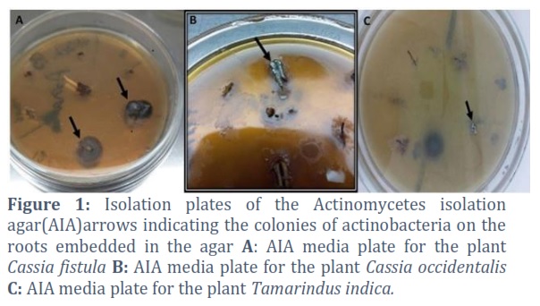
The 40 endophytic actinobacterial strains were selectively isolated from the roots of 8 different medicinal plants. Among the three different selective isolation media used, the actinobacteria colonies mainly appeared on actinomycetes isolation agar (AIA) (Figure 1). The endophytic actinobacteria n=12 was recovered from Cassia fistula, while the lowest or minimum number (n=1) of isolates was recovered from the roots of the plant Tamarindus indicia. The actinobacteria colonies appeared on the isolation agar were observed under stereomicroscope and in general the colonies with the compact, hard and embedded growth on the agar were selected. Among the 40 selected strains, a few strains seemed redundant due to the same morphology on the agar surface, however all the strains were preceded for identification and further investigation.
Morphological Characteristicsof the Selected Endophytic Actinobacteria
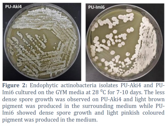
The morphology of the isolates was observed and documented after 7-10 days growth at 28 °C on the GYM agar. The growth pattern of most of the isolates varies, as some isolates were slow grower and growth appeared on the media after 8-10 days while some colonies appeared within 5 days of incubation (Figure. 2).The strains produced a variety of colored rounded to irregular colonies ranging in various size, with convex elevation, hard texture and dry or powder like aerial spores. The gram staining showed the typical gram-positive filamentous pattern in the form of chain while observed on the glass slide at 100X using oil immersion. Such as, among the strains originated from Cassia occidentalis, the strain PU-Aki2, produced off white growth on A- media with embedded colonies, the strain PU-Aki3 produced creamy off-white growth, similarly the strain PU-Aki4 produced off white regular colonies. The strains isolated from the plant Bauhinia variegata exhibited characteristic actinobacteria like features such as, the strain PU-Kach5 and the strain PU-Kach15 produced off-white growth, regular colonies. The strain isolated from Phyllanthus emblica PU-Aml3 produced creamy greenish slightly filamentous colonies. The isolate of Cymbopogon (Lemongrass) produced creamy whitish regular colonies when grown on A-media under the specified growth conditions.
Biochemical and Physiological Characteristics of the Selected Endophytic Actinobacteria
Among all the isolates 73% exhibited the production of melanin (tyrosine hydrolysis, while 80% of the isolates showed esculin hydrolysis. The utilization of various sugars as carbon source was investigated and almost all the strains showed flourishing growth on glucose, fructose, mannose, arabinose, galactose, and sucrose, while moderate and less growth was observed on rhamnose, raffinose and myo-inositol. All the isolates utilized organic acid (sodium malonate) and turned the media from greenish to blue. The enzymatic hydrolysis was positive for all isolates, almost all the strains exhibited the ability to produce cellulase, pectinase and amylase enzymes. In hemolysis test all the isolates showed transparent zones around the colonies while grown on bloods supplemented media. The whole cell analysis for DAP (Diaminopimelic acid) and carbohydrates revealed the presence of both isomers meso and L-DAP and smear of amino acid and sugars were observed on the stained TLC based on colors. The indole acetic acid (IAA) production was exhibited by all the strains as indicated by the appearance of pink color in the culture broth by adding Salkowski’s reagent except PU-Amt1, PU-Tul1 and PU-Mor2. Similarly, phosphate solubilization was observed in case of almost all the isolates, which is another indicative of the plant growth is promoting ability of the endophytic actinobacteria strains along with the IAA production.
16S rRNA Gene Sequencing of the Selected Endophytic Actinobacteria Strains
The selected isolates based on the activity and/or metabolic LCMS profiles were identified using molecular genetics approach by 16SrRNA gene sequencing and the % similarity at genus level was determined, on the basis of which a neighbor joining phylogenetic tree was constructed by using MEGA 6.0 at 1000 bootstrap replicates (Figure 3). The majority of the strains belonged to the genus Streptomyces which includes PU-Aki3 GenBank accession number; MW080716, PU-Aki4 (MZ703183), PU-Kach5 (MW080762), PU-LG1 (MW080760), PU-Imi6 (MW080759), PU-Amt17 (MW079406), PU-Amt14 (MW077167), PU-LG2 (MW080715). While the strains PU-Aki5 (MW080667) belonged to the genus Micromonospora, PU-Amt1 (MW080707) and PU-Amt9 (MW080693) belonged to the genus Nocardia.
Antimicrobial Activity of the Methanolic Crude Extracts
The crude extracts exhibited significant antimicrobial activity against the ATCC test strains by microtiter plate assay. About 53% of the methanolic extracts significantly inhibited the growth of S. aureus including the extracts of the strainsPU-Mor1, PU-Kach5, PU-Aki2, PU-Amt14, PU-LG3, PU-Imi6, PU-LG1, PU-Aki1, PU-Amt9, PU-Aml3, PU-Tul4, PU-Kach2, PU-Amt7, PU-Aml1, PU-Kach9, PU-Aki3, PU-Amt15, PU-Amt17. While moderate antimicrobial activity was exhibited by 47% of the extracts against E. coli, among which the major inhibition was observed in case of the extract of strains PU-Aki4, PU-Kach5 and PU-Amt17. The antifungal activity was exhibited by 32% of the extracts including the extracts of strains PU-Aki2, PU-LG3, PU-Amt14, PU-Kach14, PU-Aki1, PU-Tul4, PU-Amt7, PU-Kach11,PU-Kach9, PU-Kach6 and PU-Aml6 (Figure. 4).
Cytotoxicity and In-vitro Antitumor Activity of the Extracts
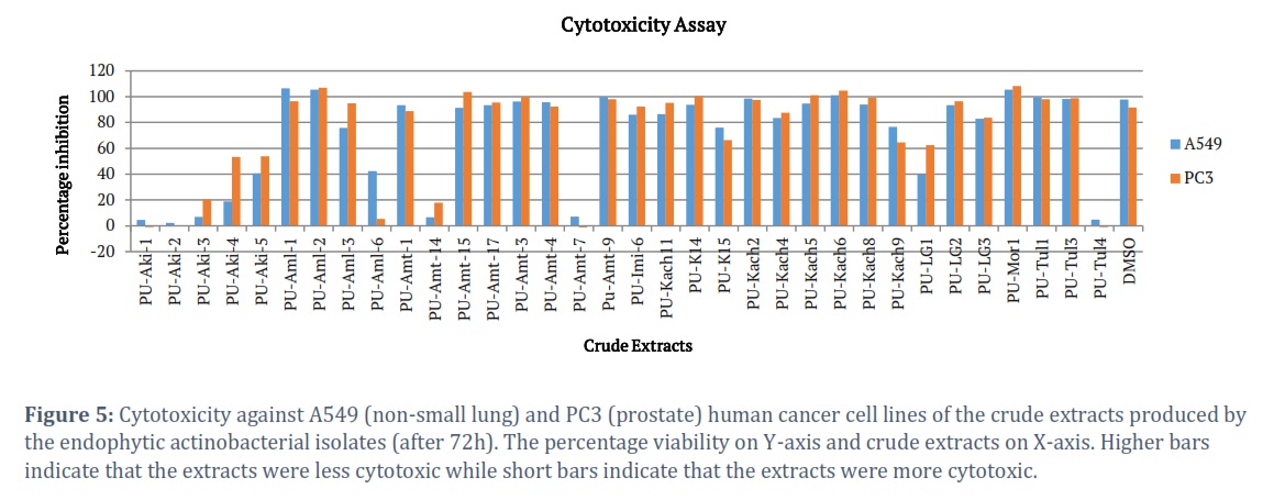
The extracts exhibited significant cytotoxicity or anti proliferative activity against the cancerous cell line PC3 (prostate cancer cell line) as compared to the A549 (non-small lungs carcinoma cell line). A total of 30% of crude extracts exhibited maximum cytotoxicity with percentage viability of cells less than 50%. The extracts of the strains PU-Aki1and PU-Aki2 exhibited 0% cell viability for both of the cell lines, the extract of strain PU-Aki3 exhibited 20% cell viability for PC3 and less than 10% viability for A549, similarly the extract of strain PU-Aki4 showed 20% viability for A549 and 50% for PC3, while the extract of strain PU-Aki5 showed 50% cell viability for both of the cell lines. The extract of strain PU-Aml6 was more cytotoxic to PC3 (10% cell viability) as compared to A549 (40% cell viability, similarly the extract of strain PU-Amt14 exhibited less than 20% viability of both of the cell lines and PU-LG1 showed about 50% cell viability (Figure 5).
DPPH Radical Scavenging Activity
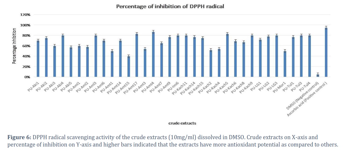
The in-vitro DPPH radical scavenging activity was exhibited by most of the crude extracts, and in most of the cases more than 50% radical scavenging activity was observed. The maximum % inhibition of DPPH was exhibited by strains PU-Aki4, PU-Kach5 and PU-Amt 17 as compared to the ascorbic acid (positive control) (Figure 6).
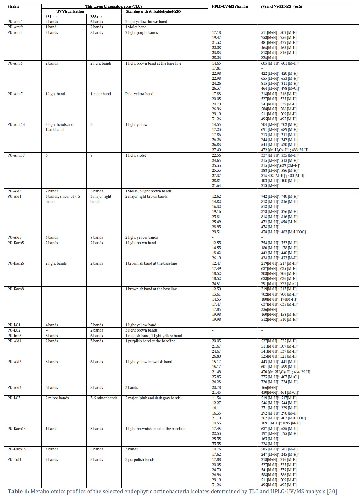
Metabolomic Profiles of the Selected Endophytic Actinobacteria Strains
The bioactive metabolites present in the extracts of the strains showed different colored bands on TLC after staining with anisaldehyde/H2SO4 reagent, some prominent bands were observed in case of the extracts of strains PU-Amt17, PU-LG1, PU-Aki2, PU-Aki4, PU-Aki5 and PU-Aml1.The fluorescent spots of red, blue, yellowish, violet and orange-color were seen under UV (366nm). A few of the bands were visualized under short UV (254nm), the prominent UV absorbing bands were observed in case of the extract of strains PU-Aki4, PU-Aki5. After staining the TLC plate with staining with anisaldehyde/H2SO4 reagent; reddish, blue, and light yellowish- and violet-colored bands were observed and were documented. The HPLC-UV/MS metabolic profile of strainPU-Aki4 displayed several bacterial metabolites eluted at different retention (tR12 ~ 29 min) with molecular masses ranges m/z = 377 ~817. The crude extracts of the strain PU-Kach5 showed 5 prominent peaks at tR12~ 27 min with molecular masses ranges m/z=179-~41. The crude extracts of the strain PU-Amt17 showed distinctive peaks eluted at retention timetR22~ 29 min, and masses of these eluted metabolites were m/z = 213 ~ 401 (Table 1).
Figures & Tables


Currently, in drug discovery, the endophytic actinobacteria are attractive candidates due to the perception that their untapped genome may encodes novel bioactive compounds, which have been proved by several studies [31-34]. So it is anticipated that the endophytic actinobacteria are a rich source of the diversified metabolites with respect to structure and function [35]. In this study 40 endophytic actinobacteria strains were isolated following stringent protocols of plant sample’s surface disinfection following themethods of Cao et al. [19]. All the isolated and subsequently selected strains showed the typical morphological characteristics of the group actinobacteria as mentioned in Bergey’s manual of bacteriology (Volume II) [20]. The biochemical and physiological characterization further confirmed that these isolates are the member of different actinobacterial genera showing the typical actinobacteria like characteristics. For example the pigment production is one of the characteristics feature of the actinobacteria and most of the strains in this study producedexo-pigments and showed the production of melanin.Similarly most of the strains showed the hydrolysis of esculin and utilization of organic acid, while in case of the utilization of the different sugars as sole carbon source [36], isolates in this study utilized most of the tested sugars except L-Rhamnose, L-Raffinose and myo-inositol. Kim et al. [37] observed the cellulase activity of the endophytic actinobacteria isolated from the roots of the plants and a similar outcome has been observed in this study and the strains exhibited cellulose activity which further confirms the origin of these actinobacteria strains from the plant tissue.Borah and Thakur [38] reported that at least one extracellular enzyme is produced by endophytic actinobacteria while in our study; most of the isolates were able to produce cellulase, amylase and pectinase enzymes. The significance of these enzymes lies in the fact that they are responsible for the entry of the endophytic isolates into the plant tissues;however the same ability of these strains can be exploited for the industrial applications and production of such commercially useful compounds.
Several actinobacteria genera have been reported to live as endophytes, however the genus Streptomyces is found to be the predominant endophyte [35, 39, 40] a similar outcome was seen in our study as all the prioritized and bioactive strains detected in this study belonged to the genus Streptomyces.However the 16SrRNA gene sequencing and subsequent BLAST analysis helped to detect the strains belonging to actinobacterial genera other than Streptomyces, such as the strain PU-Aki5 exhibited maximum similarity with the genus Micromonospora, the strains PU-Amt9 and PU-Amt1 exhibited maximum similarity with the genus Nocardia. This shows that although the Streptomyces could be the predominant genus in the endophytic actinobacteria population however members of other actinobacterial genera also invade the plant tissues and develop mutualistic relationships with the plants.
The inhibitory activity of the extracts was observed against both gram-positive and gram-negative test strains, however the prominent inhibition was observed against gram-positive bacteria especially S. aureus. Among the 40 endophytic actinobacteria strains, 18 strains originated from Cassia occidentalis and Cassia fistulashowed inhibition of S. aureus. The 10 endophytic strains exhibited significant antifungal activity while 15 were active against E. coli. In screening studies it has been observed that the activity against Gram negative test strains is usually less as compared to the activity against Gram positive test strains, as reported by Benhadjet al.[41] and Assad et al. [42]. In case of our study the response was same, the activity of the isolated endophytic actinobacteria was more prominent against gram positive bacteria. The in-vitro antitumor activity activity against human cancerous cell lines was exhibited by the extracts between moderate to high cytotoxicity against A549 and PC3 cancer cell lines, the almost similar response by endophytic actinobacteria was reported by Conti et al.[43], where the strains exhibited moderate to high cytotoxicity against the tested cell lines. In general it was observed the endophytic actinobacteria strains recovered from Cassia occidentalis showed more prominent antimicrobial and in-vitro antitumor activity or high cytotoxicity as compared to the strains retrieved from other medicinal plants investigated in this study.The production of indole acetic acid (IAA) by most of the strains indicate that the endophytic actinobacteria contribute towards the plant growth promotion (PGP) of the host plants. It supports the assumption that the endophytic actinobacteria cause positive effect on the growth of their host as already reported by Passariet al. [44].The antioxidant potential,as is evident by DPPH radical scavenging activity,show these isolates as the potential source for the isolation and discovery of antioxidant compounds,the antioxidant potential of the endophytic actinobacteria has already been reported in many studies [45].
The metabolomics profiles of the isolated endophytic strains were determined by thin layer chromatography (TLC) and HPLC-UV/MS analysis depicted the presence of variety of metabolites in the extracts of these endophytic strains. For examplenumerous UV absorbing, florescent and colored compounds were observed on TLC, the different colored compounds that appeared after staining with anisaldehyde/H2SO4 reagent shows the metabolites present in the extracts belong to different structural classes of the bioactive compounds.Similarly, a range of molecular masses of the low molecular weight compounds present in the crude extracts was detected,such as in case of strain PU-Aki4 the molecular masses of the metabolites in extracts were asfollows;m/z = 741, 817, 518, 377 and 438 Daltons, etc.while in case of strain PU-Amt17, the molecular masses of the compounds were as follow; m/z = 336, 314, 387, 401 and 213 Daltons,etc.This detected range of low molecular weight compounds with potent antimicrobial activities indicate the rich diversity of the metabolites produced by the isolated endophytic actinobacteria strains. The chemical analysis in turns further helped to prioritize the strains for media optimization and scale up studies of the priority strains in this collection.
The study revealed that the endophytic actinobacteria present in the tissues of the selected medicinal plants which are frequently used as folk medicines in south Asian region are a rich and untapped source of bioactive and structurally diversecompounds, as is indicated by their potent antimicrobial and cytotoxic/anticancer activities and distinct metabolomics profiles. Among the 40 isolated endophytic actinobacteria strains, the predominant genus was Streptomyces however members from other genera such as Micromonospora and Nocardia were also identified.The 7 strains in this collection were selected as priority strains based on their preliminary screening for scale-up fermentation, compounds purification and structure elucidation.The study shows, the continuous isolation and screening of endophytic actinobacteria from these medicinal plants may yield pharmaceutically useful metabolites and compounds.
Acknowledgment
This work was supported by Higher Education Commission (HEC) Pakistan grant (HEC-NRPU-Project 2121). The work was also supported by NIH grant R37 AI52218 (JST), the UK Center of Biomedical Research Excellence (COBRE) in Translational Chemical Biology (P20 GM130456) and the University of Kentucky College of Pharmacy and Center for Clinical and Translational Sciences (UL1 TR000117 and UL1 TR001998).The data presented in this manuscript is part of the PhD thesis of author Ashba Hassan.
Competing Interest
The authors declare that there is no conflict of interest.
![]()
References
- Bais H P, Weir T L, Perry L G, Gilroy S, Vivanco J M. The role of root exudates in rhizosphere interactions with plants and other organisms. Annual Review of Plant Biology, (2006);57:233-266.
- Liu C, Zhuang X, Yu Z, Wang Z, Wang Y, Guo X, Xiang W, Huang S. Community structures and antifungal activity of root-associated endophytic actinobacteria of healthy and diseased soybean. Microorganisms, (2019);7:243.
- Purushotham N, Jones E, Monk J, Ridgway H. Community structure, diversity and potential of endophytic bacteria in the primitive New Zealand medicinal plant Pseudowintera colorata. Plants, (2020); 9:156.
- Tian B-Y, Cao Y, Zhang K-Q. Metagenomic insights into communities, functions of endophytes and their associates with infection by root-knot nematode, Meloidogyne incognita, in tomato roots. Scientific Reports, (2015); 5:1-15.
- Silva C, Vitorino L, Mendonça M, Araújo W, Dourado M, Albuquerque L, Soares M, Souchie E. Screening of plant growth-promoting endophytic bacteria from the roots of the medicinal plant Aloe vera. South African Journal of Botany, (2020); 134:3-16.
- Sharma A, Malhotra B, Kharkwal H, Kulkarni G T,Kaushik N. Therapeutic agents from endophytes harbored in Asian medicinal plants. Phytochemistry Reviews,(2020); 19:691-720.
- Van der Meij A, Willemse J, Schneijderberg M A, Geurts R, Raaijmakers J M,van Wezel G. P. Inter-and intracellular colonization of Arabidopsis roots by endophytic actinobacteria and the impact of plant hormones on their antimicrobial activity. Antonie Van Leeuwenhoek, (2018); 111:679-690.
- Jiang Z K, Tuo L, Huang D L, Osterman I A, Tyurin A P, Liu S W,Sun C. H. Diversity, novelty, and antimicrobial activity of endophytic actinobacteria from mangrove plants in Beilun Estuary National Nature Reserve of Guangxi, China. Frontiers in Microbiology, (2018); 9: 868.
- Xu D B, Ye W W, Han Y, Deng Z X, Hong K. Natural products from mangrove actinomycetes. Marine Drugs, (2014);12(5).
- Qin S, Chen H H, Zhao G Z, Li J, Zhu W Y, Xu L H, Li W J. Abundant and diverse endophytic actinobacteria associated with medicinal plant Maytenusaustroyunnanensis in Xishuangbanna tropical rainforest revealed by culture‐dependent and culture‐independent methods. Environmental Microbiology Reports, (2012); 4(5): 522-531.
- Zhang X, Ren K,Zhang L. Screening and preliminary identification of medicinal plants endophytic actinomycetes used for inhibiting penicillin-resistant Staphylococcus aureus. International JournalofBiology, (2012); 4(2): 119–124.
- Zhang J, Wang J D, Liu C X, Yuan J H, Wang X J, Xiang W S. A new prenylated indole derivative from endophytic actinobacteria Streptomyces sp. neau-D50. Natural Product Research, (2014); 28(7): 431-437.
- Igarashi Y, Iida T, Sasaki Y et al. Isolation of actinomycetes from live plants and evaluation of antiphytopathogenic activity of their metabolites. Actinomycetologica, (2002); 16 (1): 9 – 13.
- Sasaki T, Igarashi Y, Saito N, Furumai T. TPU-0031-A and B, new antibiotics of the novobiocin group produced by Streptomyces sp. TP-A0556. The Journal of Antibiotics, (2001);54(5): 441-447.
- Sasaki T, Igarashi Y, Saito N, Furumai T. Cedarmycins A and B, new antimicrobial antibiotics from Streptomyces sp. TP-A0456. The Journal of Antibiotics, (2001); 54(7): 567-572.
- Bieber B, Nüske J, Ritzau M, Gräfe U. Alnumycin a new naphthoquinone antibiotic produced by an endophytic Streptomyces sp. The Journal of Antibiotics, (1998); 51(3): 381-382.
- Vo Q A, Ballard R A, Barnett S J, Franco C M. Isolation and characterisation of endophytic actinobacteria and their effect on the growth and nodulation of chickpea (Cicer arietinum). Plant and Soil, (2021); 466(1): 357-371.
- Wang P, Kong F, Wei J, Wang Y, Wang W, Hong K, Zhu W. Alkaloids from the mangrove-derived actinomycete Jishengellaendophytica 161111. Marine Drugs, (2014); 12(1): 477-490.
- Cao L, Qiu Z, Dai X, Tan H, Lin Y, Zhou S. Isolation of endophytic actinomycetes from roots and leaves of banana (Musa acuminata) plants and their activities against Fusarium oxysporumf. sp. cubense. World Journal of Microbiology and Biotechnology, (2004); 20(5): 501-504.
- Bergey D. H. Bergey's Manual® of Systematic Bacteriology (Vol. 2). Springer Science & Business Media, (2001).
- Shirling E B, & Gottlieb D. Cooperative description of type cultures of Streptomyces III. Additional species descriptions from first and second studies. International Journal of Systematic and Evolutionary Microbiology, (1968); 18(4): 279-392.
- Becker B, Lechevalier M P, Gordon R E, Lechevalier H A. Rapid differentiation between Nocardia and Streptomyces by paper chromatography of whole-cell hydrolysates. Applied Microbiology, (1964); 12(5): 421-423.
- Minotto E, Milagre L P, Oliveira M T,Van Der Sand S T. Enzyme characterization of endophytic actinobacteria isolated from tomato plants. Journal of Advanced Scientific Research, (2014); 5.
- Yandigeri M S, Meena K K, Singh D, Malviya N, Singh D P, Solanki M K, Arora D K. Drought-tolerant endophytic actinobacteria promote growth of wheat (Triticum aestivum) under water stress conditions. Plant Growth Regulation, (2012); 68(3), 411-420.
- Widdig M, Schleuss P M, Weig A R, Guhr A, Biederman L A, Borer E T, Spohn M. Nitrogen and phosphorus additions alter the abundance of phosphorus-solubilizing bacteria and phosphatase activity in grassland soils. Frontiers in Environmental Science, (2019); 7: 185.
- Shaaban K A, Wang X, Elshahawi S I, Ponomareva L V, Sunkara M, CopleyG C, Thorson J S. Herbimycins D–F, Ansamycin Analogues from Streptomyce s sp. RM-7-15. Journal of Natural Products, (2013); 76(9): 1619-1626.
- Wang X, Shaaban K A, Elshahawi S I, Ponomareva L V, Sunkara M, Zhang Y, Thorson J S. Frenolicins C–G, pyranonaphthoquinones from Streptomyces sp. RM-4-15. Journal of Natural Products, (2013); 76(8): 1441-1447.
- Shaaban K A, Elshahawi S I, Wang X, Horn J, Kharel M K, Leggas M, & Thorson J S. Cytotoxic indolocarbazoles from Actinomaduramelliaura ATCC 39691. Journal of Natural Products, (2015); 78(7): 1723-1729.
- Savi D C, Shaaban K A, Gos F M W R, Ponomareva L V, Thorson J S, Glienke C, Rohr J.Phaeophleospora vochysiae Savi & Glienke sp. nov. isolated from Vochysia divergens found in the Pantanal, Brazil, produces bioactive secondary metabolites. Scientific Reports, (2018); 8(1): 1-10.
- Hassan, A. (2022). Isolation and screening of endophytic actinobacteria from selected medicinal plants for antimicrobial and anti-tumor compounds [Doctoral Dissertion University of the Punjab]. Higher Education Commission Repository.1917135931.
- El-Gendy M M, El-Bondkly A M. Production and genetic improvement of a novel antimycotic agent, saadamycin, against dermatophytes and other clinical fungi from endophytic Streptomyces sp. Hedaya48. Journal of Industrial Microbiology and Biotechnology, (2010); 37:831-841.
- Pullen C, Schmitz P, Meurer K, Bamberg D D v, Lohmann S, Franca S D C, Groth I, Schlegel B, Möllmann U,Gollmick F. 2002. New and bioactive compounds from Streptomyces strains residing in the wood of Celastraceae. Planta, (2002); 216:162-167.
- Igarashi Y. Screening of novel bioactive compounds from plant-associated actinomycetes. Actinomycetologica, (2004); 18:63-66.
- Castillo U F, Strobel G A, Mullenberg K, Condron M M, Teplow D B, Folgiano V, Gallo M, Ferracane R, Mannina L,VielS. Munumbicins E-4 and E-5: novel broad-spectrum antibiotics from Streptomyces NRRL 3052. FEMS Microbiology Letters, (2006); 255:296-300.
- Qin S, Xing K, Jiang J H, Xu L H,Li WJ. Biodiversity, bioactive natural products and biotechnological potential of plant-associated endophytic actinobacteria. Applied Microbiology and Biotechnology, (2011); 89:457-473.
- Bajaj B K, Sharma P. An alkali-thermotolerant extracellular protease from a newly isolated Streptomyces sp. DP2. New Biotechnology, (2011); 28:725-732.
- Kim T U, Cho S H, Han J H, Shin Y M, Lee H B, Kim S B. Diversity and physiological properties of root endophytic actinobacteria in native herbaceous plants of Korea. The Journal of Microbiology, (2012); 50:50-57.
- Borah A,Thakur D. Phylogenetic and functional characterization of culturable endophytic actinobacteria associated with camellia spp. for growth promotion in commercial tea cultivars. Frontiers in Microbiology, (2020); 11:318.
- Tan H, Cao L, He Z, Su G, Lin B,Zhou S. Isolation of endophytic actinomycetes from different cultivars of tomato and their activities against Ralstonia solanacearum in vitro. World Journal of Microbiology and Biotechnology, (2006); 22:1275-1280.
- Taechowisan T, Peberdy J F,Lumyong S. Isolation of endophytic actinomycetes from selected plants and their antifungal activity. World Journal of Microbiology and Biotechnology, (2003); 19:381-385.
- Benhadj M, Gacemi-Kirane D, Menasria T, Guebla K, Ahmane Z. Screening of rare actinomycetes isolated from natural wetland ecosystem (Fetzara Lake, northeastern Algeria) for hydrolytic enzymes and antimicrobial activities. Journal of King Saud University-Science, (2019); 31(4): 706-712.
- Assad B M, Savi D C, Biscaia S M, Mayrhofer B F, Iantas J, Mews M, de Oliveira J C, Trindade E S, Glienke C. Endophytic actinobacteria of Hymenachneamplexicaulis from the Brazilian Pantanal wetland produce compounds with antibacterial and antitumor activities. Microbiological Research, (2021); 248:126768.
- Conti R, Chagas F O, Caraballo‐Rodriguez A M, Melo W G d P, do Nascimento A M, Cavalcanti B C, de Moraes M O, Pessoa C, Costa‐Lotufo L V, Krogh R. Endophytic actinobacteria from the Brazilian medicinal plant Lychnophora ericoides Mart. and the biological potential of their secondary metabolites. Chemistry &Biodiversity, (2016); 13:727-736.
- Passari A K, Mishra V K, Gupta V K, Yadav M K, Saikia R, Singh B P. In vitro and in vivo plant growth promoting activities and DNA fingerprinting of antagonistic endophytic actinomycetes associates with medicinal plants. PLoS One, (2015); 10(9).
- Larasati F, Batubara I, Lestari Y. The presence of endophytic actinobacteria in mangosteen peel (Garcinia mangostana) and its antioxidant activity. Biodiversitas Journal of Biological Diversity, (2020); 21(4).
This work is licensed under a Creative Commons Attribution-Non Commercial 4.0 International License. To read the copy of this license please visit: https://creativecommons.org/licenses/by-nc/4.0